DMSO et vaisseaux sanguins ( 2ème partie)
extrait de : https://www.midwesterndoctor.com/p/dmso-heals-blood-vessels-and-could?
La mise en ligne du texte par tronçons s'avère tout aussi problématique
DMSO and Blood Thinners
In medicine a balance often has to be struck between a therapeutic effect and the toxicity it entails, and getting it right can be quite challenging, such as in ensuring chemotherapy kills enough cancer cells without killing too many normal ones. Blood thinning poses another version of this problem: coagulation can predispose someone to severe complications (e.g., embolic strokes), but too little of it is also dangerous, since a minor injury can then produce a large hematoma and a blow to the head a lethal brain bleed.
For this reason, medicine typically reserves anticoagulants for those already at risk of clotting complications, as in atrial fibrillation, where the irregular rhythm lets clots form in the heart that can then travel to the brain and cause a stroke. It then tightly controls the degree of anticoagulation, doing enough to prevent the major event without raising bleeding risk any more than necessary. However, this is still far from perfect. Many individuals experience significant complications from anticoagulants each year, partly from incorrect dosing or monitoring, and partly because a perfect balance between preventing thrombosis and avoiding bleeding simply cannot always be achieved in the individual patient. All of this hence raises a few questions regarding DMSO.
First, can natural “anticoagulants” with a broader spectrum of action be used in lieu of the more potentially dangerous anticoagulants? Ultimately this is a difficult question to answer, as understandably, no clinician I know has taken the risk to attempt this in high risk situations (e.g., atrial fibrillation) and see if it suffices to stop the clotting (whereas gargantuan sums of data were needed to accurately assess the merit of pharmaceutical anticoagulants). However, in lower risk situations (e.g., preventing a heart attack over the next twenty years, or improving chronic illness symptoms resulting from excessive blood clotting), I have seen the natural alternatives perform extremely well without significant risk. Likewise, in acute situations (e.g., a heart attack or stroke), I have seen numerous cases where they (e.g., DMSO or a zeta potential restoration) resolved the incident prior to conventional options arriving. A few readers, in turn, have shared stories like quitting a problematic lifelong Eliquis prescription, noting DMSO not only prevented clots from forming but also dissolved existing ones and that “I’d be dead if DMSO didn’t work.”1 I, however, do not think this is wise, as the data simply does not exist to determine if this is appropriate to do, and as such, I instead favor the natural anti-clotting approaches in cases where it seems likely someone would benefit from blood thinning but their inherent risk of a severe clotting complication is too low to justify conventional anticoagulation.
Note: while safer than conventional anticoagulants, many of the natural anticoagulants are still not completely safe (e.g., since mass consumption of generic nattokinase⬖ came into vogue to counteract the spike protein, I have periodically come across cases of them causing a problematic bleed both within my social circle and from readers here).
Second, since DMSO potentiates the actions of pharmaceutical medications, is it safe to take with other anticoagulants? Presently, there is simply not enough data to answer this, but from everything I’ve been able to find, I lean to “yes,” with three major caveats: DMSO cannot be taken within at least two hours of the anticoagulant, coagulation must be carefully monitored to ensure excessive anticoagulation is not occurring (which should happen anyways) and IV DMSO should never be mixed with an anticoagulant in the same infusion. The limited datapoints I have to go off for this assessment are that:
•The German DMSO community, which has the most experience combining DMSO with conventional anticoagulants, considers this acceptable, citing patient reports, existing data that DMSO does not competitively inhibit clotting factors or irreversibly block platelet prostaglandins the way pharmaceutical anticoagulants do, and that in practice no meaningful changes in Quick-Wert (PT activity %) or INR values appear in patients on anticoagulants who use DMSO—although they did note DMSO can reduce the dose of aspirin (which targets platelets rather than the coagulation cascade), with multiple users “switching to 50 mg every other day and their coagulation values still being ‘as desired.’” That said, they still advise starting DMSO slowly, routinely checking coagulation values, and spacing it out from anticoagulants taken during the day.
Note: in the previously mentioned Russian study (where very limited information was provided), DMSO was reported to work synergistically with aspirin and no toxicity was mentioned from this combination.1
•DMSO–heparin combinations have a long track record of safety in topical and oral formulations. DMSO enhances heparin’s anticoagulant action and, through its membrane-transport properties, can even render heparin orally absorbable when the two are formulated together (rather than via the injection typically required for heparin). Likewise, the commercial gel Dolobene (Долобене), which combined DMSO with heparin has been widely used for decades without issues (as has Phlebolan spray, although only certain Phlebolan formulations contained heparin).
Note: Jim McCann (the inventor who brought the DMSO hematoxylin cancer therapy to Ecuador) emphasized that he had seen serious bleeding issues when IV DMSO was combined with IV heparin.
•Due to the size of the readership of this newsletter, I've had a unique window for identifying rare complications of DMSO, so over the last two years, I've kept a careful eye out for bleeding incidents linked to DMSO, of which a few reported nosebleeds after DMSO, a reader who'd used topical DMSO and then had a shoulder surgery cancelled because their PTT was 71,1 a friend on Eliquis whose small finger cut bled for two hours,1 and a reader I reached out to after learning they developed a GI bleed, which we concluded may have been linked to them taking a high oral DMSO dose concurrently with a high dose of nattokinase⬖ twice a day.
Note: conversely, many readers have also reported bleeding wounds healing with DMSO.
•The existing literature indicates at low doses (which is what can realistically be reached in the body from most uses), DMSO does not meaningfully affect standard clotting parameters at all (e.g. 1.1% did not affect clotting in a zebrafish model1 and 0.1% had no effect on human plasma PTT1). Conversely, at higher IV doses, according to one Russian review, DMSO (10–20%) lengthens clotting and bleeding time, decreases platelet aggregation and increases fibrinolysis,1 while at even higher IV doses, promotes coagulation (e.g., ≥20% precipitates fibrin and consumes fibrinogen1and ≥50% in monkeys caused instant hemolysis and fibrinogen precipitation alongside transiently shortening PTT1).
Note: due its safety, much higher doses of DMSO are routinely tested than most other agents, and as a result, toxicity often only shows up at DMSO concentrations orders of magnitude higher than most drugs. In tandem, because of how rapidly DMSO spreads through the body and dilutes, unless high IV doses are used or concentrated DMSO in ingested, it is quite difficult to reach DMSO’s toxic concentrations.
•Within the IV stem cell literature, complications are periodically reported from high dose DMSO infusions given to frail cancer patients, in the studies which looked at this, a small, transient increase in blood clotting was typically observed.1,2 Conversely, one case report exists of two elderly patients receiving IV DMSO then developing shortened prothrombin and partial-thromboplastin times (where the clinicians concluded this was likely due to quinine, indomethacin, or a phenothiazine being taken concurrently as they had not seen anything similar in comparable patients receiving IV DMSO).1 Additionally, from extensive review I was able to find three other cases exist where DMSO was linked to a problematic bleed,1,2,3 which given their rarity, makes it difficult to determine if DMSO or another variable was the causative factor.
Note: while DMSO in general helps CNS (e.g., brain) conditions, IV DMSO produces the most dramatic improvements. I initially thought this was likely due to more DMSO being able to reach the CNS, but now suspect it might be due to direct anticoagulation occurring alongside DMSO’s other effects.
•At lethal or near-lethal doses (far above ordinary use), especially concentrated IV DMSO, animal studies often show hemolysis plus pulmonary or GI hemorrhage.1Much of the hemolysis and some fluid-shift injury is osmotic and can be produced by other strongly hypertonic infusions at similar osmolar loads. DMSO’s high LD50 (toxic doses) is why those concentrations are reachable at all, as typically a different toxicity of a drug would make it lethal far below the levels needed for osmotic damage to occur.
In short, excessive bleeding on DMSO appears to be rare and when it does occur, typically mild. Nonetheless, if anticoagulants are being taken, care must be taken to ensure coagulation parameters are routinely monitored and DMSO is not directly combined with any of them.
Note: the consistent finding across decades of animal safety studies is that diluted DMSO is remarkably well-tolerated, and that its one dose-dependent liability on the blood is transient hemolysis at high intravenous concentrations. Rhesus monkeys given intravenous DMSO at 3 g/kg (as a 40% solution) daily for nine consecutive days showed no significant or lasting changes in blood chemistry, hematology, urine, or ocular, neurological, and cardiovascular parameters over four months of follow-up. Cats given intravenous DMSO every other day across ten doses maintained normal hematology and biochemistry throughout, with no cumulative or delayed adverse effects, and a DMSO-containing mixture fed chronically at up to five times the therapeutic dose produced no adverse changes (if anything, hematologic parameters improved). The primary issue repeatedly seen is that highly concentrated intravenous DMSO has been associated in older toxicity reports with local vascular irritation and transient hemolysis (e.g., 10% intravenous DMSO given to horses produced a transient dip in red-cell and hemoglobin counts that recovered within 24–48 hours).
Effects on Prostaglandins & Thromboxane
Prostaglandins and thromboxane are the short-lived signaling molecules the body makes from arachidonic acid (via the cyclooxygenase, or COX, enzymes) to control clotting, vascular tone, and inflammation. The system has two opposing sides, thromboxane A₂ (TXA₂), which drives platelet aggregation and vasoconstriction and prostacyclin (PGI₂) and PGE₁, which do the reverse, dispersing platelets and dilating vessels. As such, the balance of these heavily influences whether blood clots and vessels constrict, or blood flows and vessels open.
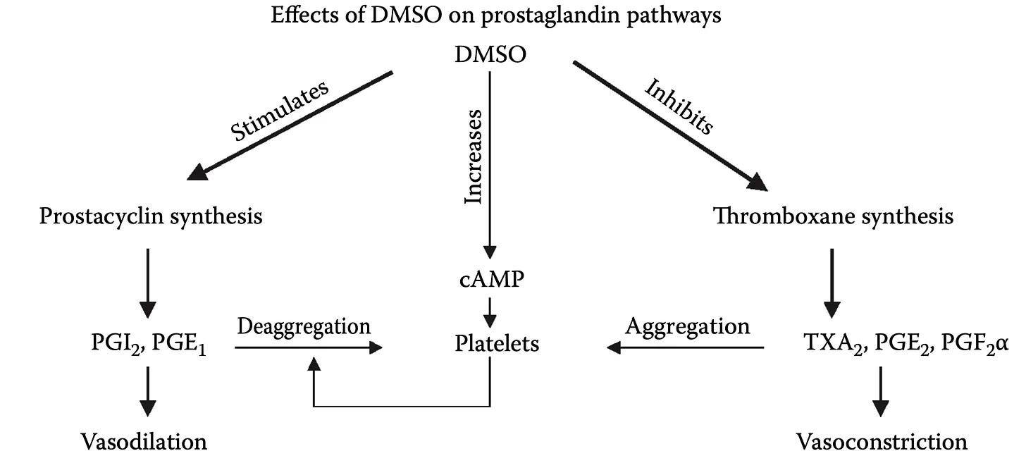
DMSO is often described as a prostaglandin inhibitor (an aspirin-like agent that blocks COX) as for platelets, DMSO selectively inhibits COX-1 and thromboxane A₂ synthase,1,2 cutting TXA₂ production and in cholesterol-fed rabbits, DMSO lowered arterial atherosclerosis, an effect attributed to its reduction in platelet thromboxane B₂ (a stable marker of TXA₂) while prostacyclin was untouched.1 So, since DMSO raises PGE₁ and cAMP while decreasing thromboxane and fibrinogen,1 it shifts the balance towards reducing clotting and dilating vessels.
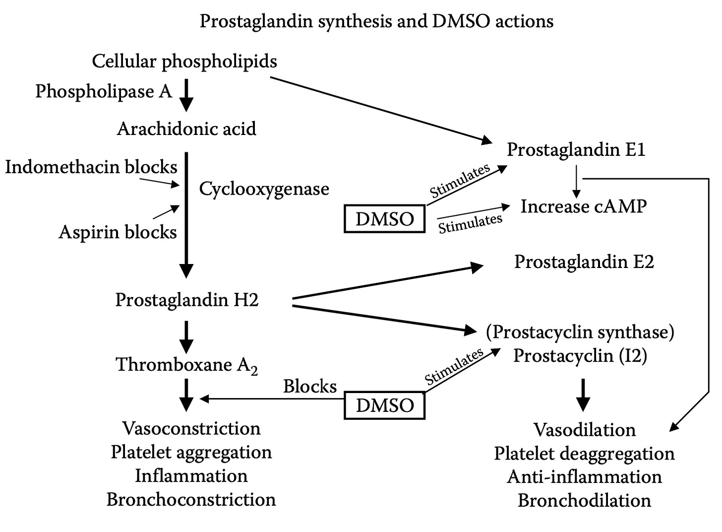
Note: the fuller picture is more complex than “DMSO blocks COX,” as data shows DMSO is not uniformly a prostaglandin-synthesis inhibitor. In one study it increased arachidonic-acid oxidation rather than inhibiting it, didn’t suppress thromboxane, and led its authors to propose it acts as a reducing cofactor rather than a blocker.1 Another found DMSO had no effect on thromboxane yet stimulated PGE₂ and concluded DMSO’s anti-inflammatory action doesn’t run through an arachidonic-acid metabolism at all.1 On skin, DMSO releases prostaglandins and arachidonic acid into the tissue1 (much as histamine does), while in cultured aortic endothelial cells it actually inhibited prostacyclin.1 All of this argues that DMSO doesn’t switch the eicosanoid system off but rather rebalances it, with the net effect in platelets and vessels tilting toward the antithrombotic, vasodilatory side (consistent with DMSO’s broader tendency to normalize dysregulated systems rather than force them one way). Likewise, as we’ve seen before, the effect is also context-dependent, as in some models DMSO adds nothing, and did not improve sevoflurane’s cardioprotection when tested on its own.
Finally, DMSO has also been used to therapeutically deliver beneficial prostaglandins. In one report, topical PGD2 combined with DMSO was used to save digits (fingers or toes) that would otherwise be lost from acute inoperable thrombosis,1 and has been used to deliver agents that act upon this system (e.g., tetrahydroxystilbene⬖1 and curcumin⬖1 to relax the superior mesenteric arteries and arterioles).
Raising cAMP & cGMP
Two closely related molecules cyclic AMP (cAMP) and cyclic GMP (cGMP), once elevated, in platelets, reduce their aggregation and in vascular smooth muscle trigger vessel relaxation and widening.
DMSO raises cAMP through both increasing its production and by slowing its breakdown. On one hand, DMSO directly stimulates adenylate cyclase,1,2,3 the enzyme that synthesizes cAMP; in heart tissue this stimulation was maximal at around 10% DMSO, comparable in magnitude to the beta-agonist isoproterenol and additive with it—yet DMSO’s effect was unaffected by α- or β-adrenergic blockade, meaning it works independently of the adrenergic receptors those drugs act on.1 DMSO also was found to inhibit cyclic AMP phosphodiesterase1 (the enzyme that degrades cAMP), which would further raise cAMP levels.
As such, in platelets DMSO significantly raises both cAMP and cGMP (through a channel entirely separate from its COX-1 inhibition), further supporting its antiplatelet effects.1 In blood vessels, DMSO likewise raises cGMP and relaxes the vessel wall through a nitric-oxide/cGMP-dependent pathway.1
Note: like DMSO’s other effects, this one is concentration-dependent and not universal—at lower concentrations or in certain tissues DMSO showed no measurable effect on the cyclase system.1,2,3
Opening Blood Vessels (Vasodilation)
Blood flow is controlled by smooth muscle surrounding the lining (endothelium) of the blood vessels, which tightens to restrict flow. As such, DMSO’s ability to relax that muscle provides another key way for it to restore tissue circulation. DMSO’s circulatory-promoting properties turn up across nearly every vascular bed studied (e.g., aorta, coronary, pulmonary, mesenteric, renal, cerebral, umbilical, and saphenous) both relaxing vessels by itself and serving as the medium through which other vasodilators are delivered without impeding their effect.1,2,3,4
In the aorta, DMSO produced a concentration-dependent relaxation of contracted aortic rings through an endothelium-dependent pathway (via nitric oxide and cGMP), and an endothelium-independent one that inhibits calcium channels in the smooth muscle and reduces the muscle’s sensitivity to calcium (partly via Rho-kinase).1,2Likewise, in another study, DMSO relaxed aortic rings by 42-99% and renal arteries by 80% (in part through voltage-gated potassium channels),1 in another DMSO relaxed mesenteric artery preparations, and in drug references, vasodilation is one of dimexide’s (DMSO) recognized pharmacological properties.1
Note: since one of DMSO’s relaxing effects acts directly on smooth muscle, it likely can work on damaged vessels where the endothelium is no longer responsive to stimuli.
Additionally, part of DMSO’s local vasodilation is histamine-mediated—the same mechanism behind the warmth and flushing people often feel where DMSO is applied. Importantly, this is a controlled dilation rather than tissue injury, as when DMSO was injected into a limb, it markedly increased lymph flow while the protein content of that lymph fell (the opposite of what happens with a burn or a caustic agent, where protein rises) and tissue examination showed only vasodilation and occasional edema, but no cell necrosis (death).1 DMSO hence opens vessels and shifts fluid without damaging the tissue.
Note: combined with hydrogen peroxide as an oxygen source, topical DMSO raised skin-flap survival to 92% versus 71% in controls, an effect the authors attributed partly to DMSO’s vasodilatory, histamine-like action.1
DMSO and Nitric Oxide
The classic way a blood vessel dilates is that the endothelium produces nitric oxide, which diffuses into the surrounding smooth muscle and triggers relaxation there through cGMP. For this reason, many integrative cardiovascular health approaches focus on supporting endothelial nitric oxide production.
Much of DMSO’s own vasorelaxation runs through the nitric oxide/cGMP pathway as in isolated aorta, DMSO’s relaxation is blunted by removing the endothelium or by blocking nitric oxide synthase with L-NAME, while conversely, DMSO raises tissue cGMP the downstream marker of nitric oxide signaling1 and at 10% redistributed intracellular nitric oxide into mobile membrane vesicles.1
Furthermore, since DMSO effectively scavenges reactive oxygen species (which cripple the endothelium’s ability to make and use nitric oxide), DMSO is able to preserve the ability of blood vessels to dilate (e.g., protected against cigarette-smoke-induced impairment of nitric oxide production and vasodilation in rabbit aortas and human endothelial cells)1 and, from the opposite direction, DMSO's radical scavenging restored an endothelium-dependent, nitric-oxide–mediated relaxation that iron had blocked in aortic rings.1
Simultaneously, DMSO can counteract nitric oxide when it’s excessive, both by scavenging it directly (DMSO scavenges radicals including nitric oxide), as when IV DMSO in horses modestly raised systolic blood pressure,1 and by throttling its production, as when 2.5–3.5% DMSO (but not 1%) cut endothelial nitric oxide output1—an effect reversed by adding extra arginine (a common supplement used to enhance nitric oxide production).
Collectively, this suggests that DMSO normalizes nitric oxide function, both by preserving it when it would otherwise be impaired, but also by counteracting it if it becomes excessive and harmful (as at low doses, DMSO typically does not affect nitric oxide).
Finally, DMSO has also been used to deliver agents which preserved or restored nitric oxide mediated relaxation such as resveratrol,⬖1,2 hesperidin,⬖ (reducing hyperhomocysteinemia-induced cognitive deficits),1 thymoquinone,⬖1 red-clover isoflavones,⬖1 genistein, ⬖1,2,3,4 (e.g, for fructose-fed hypertension1) methylated quercetin flavonoids and phenylbutanoids,⬖1 Bacopa monnieri flavonoids and saponins,⬖1 tetrahydroxystilbene glucoside,⬖1 astragaloside IV,⬖1 cinnamaldehyde,⬖1Jasminum sambac extract,⬖1 ursolic acid,⬖1 Acer okamotoanum sap,⬖1 phloridzin,⬖1adenosine,⬖1 a Periplaneta americana metabolite,⬖1 a polyphenol nutraceutical,⬖1 the soluble guanylate-cyclase activator cinaciguat,1 the PPAR-γ agonist rosiglitazone,117β-estradiol,1,2 a STAT3 inhibitor,1 a selective adenosine kinase inhibitor,1 a PPAR-α agonist,1 and an angiotensin peptide analog.1
Counteracting Oxidative Constriction
A large body of literature shows DMSO’s free-radical scavenging (discussed below) protects a wide range of blood vessels from the constriction and endothelial dysfunction caused by oxidative stress, hence keeping vessels from closing down:
In the aorta, it nearly completely prevented ROS-induced vasoconstriction and preserved endothelial and contractile function1 and attenuated hydroxyl-radical- and H₂O₂-induced contractions,1 and it protected platelet-mediated, endothelium-dependent relaxation from hydroxyl-radical damage.1
In coronary arteries, it protected endothelium-dependent relaxation from radical-induced dysfunction,1 and it preserved CGRP-mediated neurogenic relaxation of arteries under oxidative attack.1
In pulmonary and cerebral vessels, it reduced hypoxic pulmonary vasoconstriction by lowering oxygen-radical release,1 and, as a reducing agent, inhibited and reversed the vasoconstriction that oxidizing agents such as peroxide and silver nitrate induce in rat aortas and dog basilar arteries.1,2
Lastly, DMSO also shielded endothelial cells from the injury driven by infection and its toxins, including bacterial endotoxins1 and Pseudomonas aeruginosa1 products.
Note: DMSO’s direct vasorelaxation is concentration-dependent so at the higher concentrations DMSO reaches its own clear relaxing effect, but at the low concentrations used in many solvent experiments, it frequently shows no measurable effect on vascular tone.1,2,3
Free Radical Scavenging
Free radicals are atoms or molecules that contain one or more unpaired electrons, which makes them highly reactive and potentially damaging to tissues such as fats, proteins, and DNA. DMSO’s most well-recognized property is its ability to scavenge (neutralize) free radicals—particularly the hydroxyl radical, one of the most damaging reactive species, of which DMSO is among the best-known scavengers (there are 135,000 results on Google Scholar for “DMSO radical scavenger”). Its reaction with the hydroxyl radical, in fact, is so reliable that DMSO is used as a standard molecular probe for detecting and quantifying hydroxyl radicals in biological systems as the hydroxyl converts DMSO into a stable, easily measured marker.1,2
Note: two of the most commonly observed (and studied) scavenging effects of DMSO are its reduction of free-radical-induced lipid peroxidation and its preservation of the body’s own antioxidant enzymes.1 Additionally, DMSO’s radical scavenging is dose dependent,1 can directly suppress hydroxyl-radical production in solution,1 and delivers vascular protection other approaches cannot—in a cobra-venom model of complement-driven vascular injury, it prevented the microvascular damage (capillary congestion and leukocyte plugging) that cyclooxygenase inhibitors, an iron chelator, and a thromboxane synthetase inhibitor all failed to touch.1
Since the thin lining of the blood vessels (the endothelium) is highly vulnerable to oxidative damage, DMSO’s ability to scavenge radicals has shown a variety of key therapeutic effects including:
•Preventing endothelial dysfunction and preserving vessel dilation.1
•Protecting aortic endothelial cells from radical-triggered programmed death.1
•Blocking the reactive-oxygen signaling that switches on the adhesion molecules (ICAM-1, VCAM-1, E-selectin) endothelial cells use to catch and recruit inflammatory white cells (detailed here) largely by inhibiting the master inflammatory switch NF-κB—thereby preventing those white cells from sticking to the vessel wall, plugging the smallest vessels and causing the microstrokes which underlie many illnesses (e.g., vaccine injuries).
•Blocking the radical-driven stiffening of endothelial cells that would otherwise impair blood flow.1
As such, many studies have shown DMSO protects the endothelium from oxidative stress and free radical injury.1,2,3,4,5,6,7,8,9,10, 11,12,13,14,15,16,17,18,19,20, 21,22,23,24,25,26,27,28,29,30, 31,32,33,34,35,36,37,38,39,40, 41,42,43,44,45,46,47,48,49,50, 51,52,53,54,55,56,57,58,59,60, 61,62,63,64,65,66,67,68,69,70, 71,72,73,74,75,76,77,78,79,80, 81,82,83,84,85,86,87,88,89,90, 91,92,93,94,95,96,97,98
In addition to neutralizing the free radicals that constrict vessels and injure the vessel wall, DMSO protects against a second wave of radical damage: when a vessel’s blood flow is cut off and then restored, the oxygen-starved (ischemic) tissue converts the returning oxygen into a burst of radicals, creating a “reperfusion injury” that often exceeds the damage from the initial blood loss. As such, in virtually every part of the body (particularly the central nervous system), DMSO has been repeatedly shown to protect organs and tissue from oxidative stress and otherwise devastating reperfusion injuries.1,2,3
For example, DMSO:
•Significantly reduced the microcirculatory vascular leakage caused by a radical-generating system in hamster cheek pouches.1
•Reduced vascular permeability and edema in thermally burned skin.1
•Protected dermal microvessels from burn injury.1
•Reduced vascular permeability, edema, and tissue injury in gut and mesenteric ischemia-reperfusion, performing comparably to the antioxidant enzymes superoxide dismutase and catalase.1,2,3,4,5,6,7,8
•Reduced damage in hemorrhagic shock and ischemic gastric injury.1,2,3,4,5,6,7,8,9,10
•Protected the lung and pulmonary vasculature from radical injury.1,2
•Prevented the secondary lung injury that radicals inflict on distant organs after a period of hind-limb ischemia.1
•Reduced endotoxin- and shock-driven organ injury.1
•Improved skin-flap and free-tissue survival by blunting reperfusion radical injury.1,2
•Protected the liver during cold-ischemic preservation.1,2
•Used to counteract the reperfusion radical injury implicated in equine laminitis.1,2,3,4
Note: in many cases, these protective effects are dose-dependent (e.g., in a rat limb ischemia-reperfusion the dose used only gave partial protection,1 while in a lung toxin model, a fairly high dose was needed to provide protection1).


Commentaires
Enregistrer un commentaire