DMSO et vaisseaux sanguins (3ème partie)
extrait de : https://www.midwesterndoctor.com/p/dmso-heals-blood-vessels-and-could?
Protecting the Endothelium
The innermost surface of the endothelium is coated with the glycocalyx, a fragile, sugar-rich gel layer that regulates permeability, shields the cells from shear, and keeps clotting and inflammation in check. In shock, sepsis, and reperfusion, this layer is stripped away (”shed”), and its loss is now recognized as an early, pivotal event in vascular collapse.
Note: sulfates are one of the most effective biological molecules for restoring (vital) zeta potential. The glycocalyx is structured so that abundant sulfates on it form a (continually regenerating) liquid crystalline gel around the glycocalyx which repels other substances from entering the endothelium and independently drives blood circulation (all of which is detailed here)—making the glycocalyx one of the most critical components of cardiovascular health.
Many studies, in turn, show DMSO protects the glycocalyx along with the underlying endothelium:
•In a mouse model of LPS-induced acute respiratory distress, systemic DMSO preserved the pulmonary endothelial glycocalyx (staining intensity roughly four times that of untreated injury), cut protein leak into the airspaces, and reduced inflammatory cell counts; in human endothelial cells it prevented glycocalyx shedding by blocking the enzyme (a matrix metalloproteinase) that cleaves it from the cell surface.1
•In a rat hemorrhage-resuscitation model, DMSO reduced glycocalyx shedding and the accompanying coagulopathy.1
•At the level of the underlying matrix, DMSO restored the normal structure of heparan-sulfate proteoglycan in endotoxin-exposed endothelial cells,1 and prevented endotoxin-induced lung protein leak while preserving the endothelium’s ability to relax.1
•DMSO also inhibited apoptosis in nutrient-deprived human endothelial cells by promoting DNA replication and survival,1 and protected them under oxidative stress by raising heme oxygenase-1 and reducing apoptosis through several cytoprotective pathways.1
•In the equine ascending colon, DMSO protected against the capillary permeability changes caused by ischemia-reperfusion injury.1
Note: DMSO has also been used in combination with a variety of agents to promote endothelial growth and repair (e.g., in studies of endothelial proliferation, migration, tube formation, differentiation, and progenitor-cell-driven revascularization). Agents combined with DMSO to create these effects on the endothelium include: curcumin, ⬖1 resveratrol,⬖1luteolin,⬖1 kaempferol,⬖1 diosgenin,⬖1 icariin,⬖1 arnebin-1,⬖1 celastrol,⬖1 dihydroartemisinin,⬖1alkannin,⬖1 sulforaphane,⬖1 ginsenoside Rg3,⬖1 Illicium henryi extract,⬖1 dracorhodin,⬖1onychin,⬖1 Echinacea extract,⬖1 13-cis retinoic acid,1 fluvastatin,1 simvastatin,1 atorvastatin,1dihydrotestosterone,1 recombinant proteoglycan-4,1 sirolimus1 rapamycin,1 a demethylating agent,1 a Kir2.1 blocker,1 or a JAK2 inhibitor.1
Current Stroke Management
Roughly 3.1% of adult Americans have experienced a stroke (a figure I expect to rise from the COVID-19 vaccines). Each year, this translates to about 800,000 people in the United States having a stroke, and in 2022, 165,393 died (making it the fifth most frequent cause of death in the United States), with between 20-40% of survivors experiencing long term disability from the stroke.
Because of the harm strokes pose to society, and the rate at which brain tissue deteriorates once its blood supply is lost, the medical system emphasizes doing everything that can be done to treat strokes as soon as possible. Unfortunately, there is a fundamental limitation to how strokes are addressed which makes it impossible to ever eliminate the immense toll strokes place upon society.
This is because three types of strokes exist, ischemic strokes (which are typically caused by a blood clot obstructing the artery), a brain hemorrhage (where bleeding is both directly toxic to sensitive brain tissue and creates large blood pockets that compress and damage the brain), and transient ischemic attacks (TIAs)—which are not classically considered strokes.
Note: TIAs occur when someone shows clinical signs of a stroke that resolves on its own (and typically does not have stroke imaging findings). I believe TIAs often represent microstrokes occurring (as the microclots are too small to be seen with conventional brain imaging and typically resolve on their own)—a subject I went into much more detail here as these visually detectable microstrokes frequently underlies vaccine injuries and many other chronic illnesses.
The essential challenge with managing strokes is that it’s nearly impossible to reliably differentiate an ischemic stroke from a hemorrhagic stroke without imaging (we’ve tried for a longtime, and while there are a few diagnostic signs that suggest one or the other, they are not reliable enough for the degree of certainty required). This matters because opposite approaches are taken to ischemic strokes and hemorrhagic strokes—with ischemic strokes a powerful clot busting medicine (tPA) is used to restore circulation, whereas with hemorrhagic strokes (13% of strokes in the developed world), a variety of steps are taken to reduce further bleeding in the brain (which can include brain surgery).
As such, when a stroke occurs, while it is urgent to treat it as soon as possible (as “time is brain”), the standard approach (tPA) cannot be used until a CT scan has ruled out a hemorrhagic stroke (as giving tPA for a hemorrhagic stroke is devastating or lethal). Because of this, definitive stroke treatment cannot begin until it has been recognized, an ambulance gets them to an ER, a CT scan has been completed, a diagnosis is made, and that diagnosis reaches the treating physician—all of which, despite the best efforts by the medical system to expedite the process, often takes hours.
Worse still, the statistics on tPA (approved in 1996 and still the only FDA approved treatment for ischemic strokes) aren’t actually that good. Presently, tPA is only approved to be given within 3 hours of a stroke starting (as its likelihood of benefitting a patient decreases with time), and in practice, it is often given up to 4.5 hours after symptoms start (since some degree of benefit still exists).
When that window is met (which only happens about 25% of the time and ultimately results in roughly 1.8%-8.5% of ischemic stroke patients receiving tPA), the existing data shows that only 13% percent of patients who receive tPA significantly benefit from it (39% return to normal, compared to 26% who would return to normal without treatment), with an additional 19% of tPA users experiencing some degree of improvement (but not a full recovery) from it.
Worse still, tPA can cause significant bleeding, which is sometimes minor (e.g., gum bleeding), but also carries a 6.4% risk of a symptomatic brain bleed, and a 1.6% risk of a serious systemic hemorrhage (along with other issues such as a 1.3% to 5.1% risk of angioedema and tPA frequently causing reperfusion injuries). In turn, many risk factors exist for the increased bleeding (e.g., a few common risk factors can lead to a 33% chance of tPA causing a fatal bleed), and there have been many lawsuits for either giving or not giving tPA to a stroke patient. Additionally, tPA is a poor choice for larger obstructions (e.g., one within the internal carotid artery), which instead must be physically removed. In short—many ICU doctors I know are quite hesitant to use tPA as they have seen cases where it dramatically improved patients, many where it did not do anything, and quite a few disasters (especially in the early days of the therapy where it was used for heart attacks and then often caused the patient to have a fatal or debilitating brain bleed).
Note: the best data exists for tPA being injected directly into the obstructed artery with interventional radiology. Unfortunately, while many premier institutions offer this, it is a specialized procedure that is not available at most hospitals.
Finally, there is essentially no therapy for recovery from stroke—which in short explains why stroke is the second leading cause of death and the third leading cause of disability worldwide.
DMSO and Stroke Management
As the previous section shows, there are insurmountable limitations to how strokes can be managed. DMSO, however, completely changes this equation as:
•It effectively treats ischemic strokes.
•It partially treats hemorrhagic strokes.
•It carries no known risk of worsening a hemorrhagic stroke.
•It can be easily administered at home or on an ambulance.
•In addition to addressing the circulatory issues, independently protects brain tissue from stroke damage.
•Prevents the reperfusion injuries which follow strokes.
•Can heal damaged brain tissue after a stroke.
As such, DMSO makes it possible to eliminate the multi-hour delay typically required for stroke treatment, often is a safer can be a more effective stroke treatment option than the conventional alternatives, and provides one of the only existing options to heal the permanent disability that follows a stroke.
This is why researchers like Jack de la Torre MD devoted their entire careers to producing a robust body of evidence DMSO could treat strokes, as they knew it was unlikely any other agent would ever be able to solve the ischemic-hemorrhagic stroke dilemma.
Note: many readers have reported to me successfully treating strokes at home or on the way to the ER with DMSO and hence, due to increasing skepticism towards hospital care,1 have advocated for home DMSO in lieu of going to the hospital. This is extremely unwise, as if a significant hemorrhagic stroke is occurring, DMSO cannot work as the primary treatment. Rather, the correct course is to use DMSO on the way to the hospital, and then if the whole thing is written off as a TIA (which many people have shared happened to them after they used DMSO, it resolved the stroke midway to the ER, and ER could then not decide if a stroke had happened), simply be grateful you had the optimal outcome when things could have easily been much worse. In short, the existing management we have for strokes is correct, and DMSO should be viewed as way to improve the results it yields rather than a replacement for it.
Ischemic Stroke Data
Many stories like these two Archie Scott reported1 exist throughout the DMSO literature:
A Los Angeles school teacher had a major stroke shortly after the start of the Christmas break. She was unconscious on her living room floor. DMSO treatment was started immediately after the stroke. The DMSO was first applied topically to her head within minutes of the stroke. Less than one hour after the stroke she was given DMSO by intramuscular injection. This patient was never taken to the hospital for this stroke. A prominent surgeon who was a family friend told the husband of this patient that it was important to keep her out of the hospital. The surgeon said that even though the treatment was completely legal, it would be difficult to get approval to give the DMSO especially by injection at his hospital.
This patient made a dramatic recovery. She regained consciousness later in the day in which she had her stroke. Treatment continued for the next week. Each day she received two topical applications of DMSO, one intramuscular injection of DMSO, and two doses of one teaspoonful of DMSO in juice. Her condition improved each day. When school resumed after the first of January, this teacher was back in the school teaching the students as if nothing had happened during the Christmas vacation. She continued teaching until she retired, healthy and with no disability.
A lady was in a coma in a convalescent hospital and had been in the coma since her stroke three months ago. She was given little chance of recovery and was expected to remain in a vegetative state until her death.
When I first observed this lady, there was no response to any type of stimulus. She was alive, but appeared lifeless. It was decided that her treatment should be topical DMSO daily….One month after the start of treatment, there were positive signs in the lady. Her brain was starting to respond to the DMSO. The treatment continued, and four months after treatment started this lady was able to return to her home [where treatment continued].
Three years after the start of DMSO treatment this writer returned to visit this patient. At this time the lady was living a normal life, not the life of a stroke victim. She was able to look after the house and walked normally.
The only lingering effect of the stroke was a slight speech defect. At this time she said that her memory was better than that of her husband who had not had a stroke and who was considered to be completely normal.
Note: there are also many reported cases of individuals who took DMSO for musculoskeletal or pain disorders (by far the most common use of DMSO) who then experienced a permanent improvement of stroke symptoms.
Extensive data, in turn, corroborates these remarkable stories.
Human Evidence
Due to the ethical issues with creating and then treating strokes in humans, most of the research in this field has been conducted in animals (which I hold many strong ethical objections to). As such, the only human study has been conducted in ischemic strokes was a 2002 trial, where IV DMSO given with fructose diphosphate twice daily to 11 patients (mean age 65) with acute or subacute ischemic stroke was well tolerated and, when started within 12 hours of stroke onset, left 63% “improved” or “markedly improved” versus 20% of those on standard care at three months, and the benefit held even when treatment began well after the stroke.
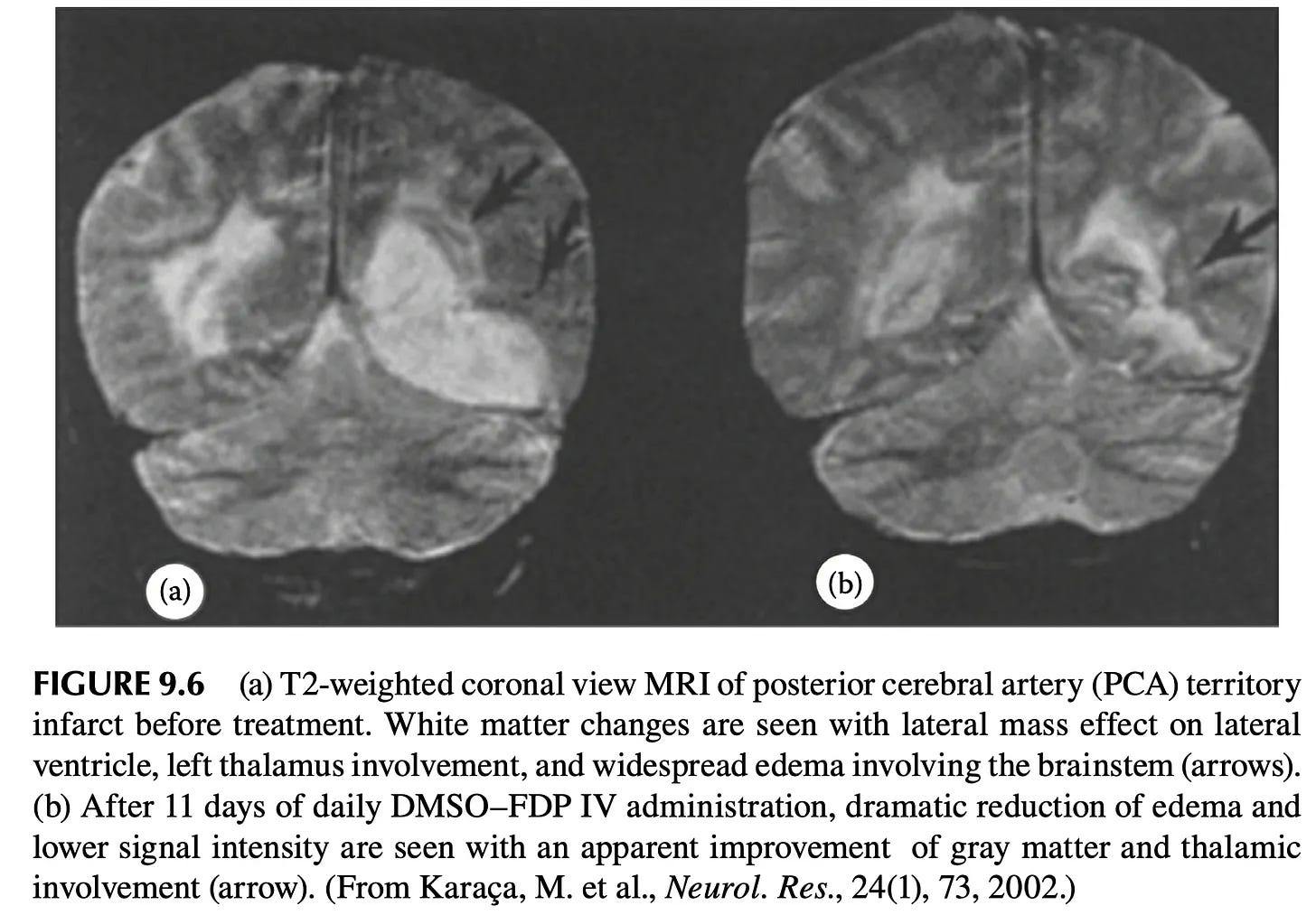
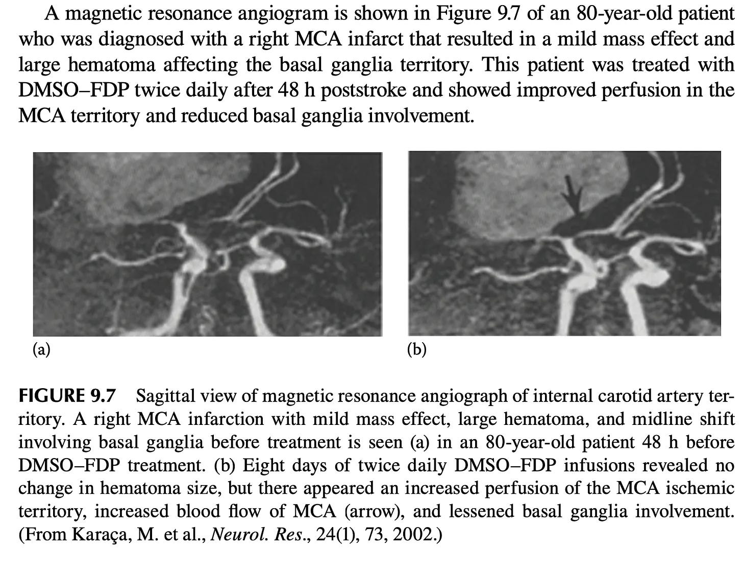
Note: animal studies further corroborate DMSO-FDP value. In rabbits whose brains were driven to flatlined (isoelectric) EEGs by combined hypoxemia, hypotension, and carotid occlusion, DMSO-FDP given after five minutes of electrical silence restored brain activity far faster and let all animals survive with minimal damage, versus only 22% survival (severely disabled) on saline, and in mice given head impacts (another area DMSO helps), DMSO-FDP was the most protective and DMSO alone the second, while FDP and the rest gave no benefit, confirming DMSO was therapeutic. Finally, when DMSO-FDP was given to rats with poor chronic blood flow to the brain (due to carotid obstruction), there was a 54% improvement in visuo-spatial memory1,2 (which is corroborated by dementia patients who’ve improved from DMSO).
•A Russian patent treated perinatal hypoxic encephalopathy in newborns by daily electrophoresis of vitamin E⬖ in DMSO. In three term infants 6–7 sessions produced disappearance of limb tremor (chin tremor remaining only with crying), normalization of muscle tone, return of tendon and support/automatic-walking reflexes, loss of the spontaneous Moro reflex, and restoration of the Babkin reflex, with faster clinical recovery than intramuscular vitamin E⬖ plus standard care.
•One physician reported knowing of a patient of Stanley Jacob’s whose stroke was successfully treated with DMSO.1
Note: I have corroborated through multiple sources that Stanley Jacob successfully treated numerous strokes with DMSO, but I have not been able quantify how many in total were treated or what the success rate was.
Additionally, numerous reviews support DMSO’s use in humans with strokes. These include:
•A 2009 pharmacology review by Jack de la Torre and Stanley Jacob of DMSO in cardiac and CNS damage (arguably the most detailed one in print) showed intravenous DMSO elevated cerebral blood flow, limited infarct volume after middle-cerebral-artery occlusion, preserved the vulnerable CA1 and dentate neurons and facilitated recovery from acute or chronic cerebral ischemia.1
Note: de la Torre was publishing papers on DMSO treating strokes fifty years ago1 and likewise, a 1982 paper1 acknowledged DMSO was being used to treat strokes.
•A 1992 Chinese review that concluded DMSO protects ischemic brain tissue and blocks the secondary pathological processes that follow cerebral ischemia, outperforming mannitol, dexamethasone, and barbiturates as a neuroprotectant.1
•A 2010 Chinese review of DMSO’s effects on the heart and central nervous system (which noted authors had identified over 10,000 articles on the biological effects of DMSO).1
•A 2012 Chinese review classified DMSO as a classic antioxidant with therapeutic effects on ischemia reperfusion injury across many organs (and noted it had already been used clinically abroad for cerebral reperfusion injury).1
•In a 2018 Ukrainian physical-rehabilitation review where DMSO appears as an iontophoresis solvent in post-stroke and disability rehabilitation protocols.1
•A 2022 Russian review from Osh State University argued that drug electrophoresis (driving charged medication ions through the skin with direct current) is among the most valuable physiotherapy methods for cerebrovascular disease (stroke and TIA) and peripheral-nerve disorders. Within it, DMSO is used as a carrier that lets otherwise water-insoluble drugs dissociate and be pushed through the skin. In acute stroke in the early recovery period, this was applied to the eyelids and back of the neck with heparin added for ischemic stroke and iodine substituted for hemorrhagic stroke.1
DMSO Alone in Stroke Models
DMSO has repeatedly been shown to reduce the core injury of an ischemic stroke:
•A rhesus monkey study blocked the MCA for 4 hours, gave DMSO, dexamethasone, or nothing, and then opened the MCA after it had been blocked for 17 hours. DMSO gave significant protection from the severe neurological deficits and loss of arterial blood flow the other two groups developed.

•A squirrel monkey study blocked the left MCA for 4 hours, after which the animals were given a variety of different treatments (e.g., saline, hemodilution, or hyperbaric oxygen at 2 atmospheres). Seven days after treatment, 8 of 10 DMSO treated monkeys were alive (with 2 having mild contralateral muscle weakness), while 75% of those receiving hyperbaric oxygen survived, and just 34% of those receiving hemodilution survived (with the last two groups also having more significant neurological deficits). Finally, combining either of these treatments with DMSO produced slightly worse results than just DMSO alone.
•In experimental canine middle cerebral artery embolectomy, DMSO (or low-dose methylprednisolone) extended the grace period to 6 hours following embolization and protected the cerebral tissue from the injury of ischemia and post-ischemic reperfusion (with no infarcted tissue in DMSO treated animals and a 1.45 cm3 area of infarction in untreated ones).1,2,3,4
•In a canine model of severe cerebral ischemia (where blood flow was cut by 90% for an hour), DMSO given alone as pretreatment allowed EEG activity to re-emerge after recirculation where untreated animals showed none. In a second canine study, in brains subjected to 14 minutes of complete ischemia and then reperfused for an hour, DMSO added to the reperfusion blood lowered a marker of lipid peroxidation (TBAR) back to control levels, restored roughly three quarters of the lost energy stores (ATP and creatine phosphate), cut lactate accumulation by 60%, and produced a marked return of EEG and auditory evoked potentials that untreated brains entirely lacked. Adding a platelet-activating factor antagonist on top of the DMSO further improved mitochondrial energy markers, though the reduction in lipid peroxidation was attributable to DMSO alone.
Note: the first study also found vitamin E⬖ restored brain function, mirroring forgotten Russian research on how to counteract the biological consequences of chronic stress on the brain with DMSO and vitamin E.⬖
•In a canine model of pressure-induced focal ischemia worsened by ethanol exposure (done to model head trauma from a drunk driving car accident), DMSO significantly reduced brain lesion volumes under both normotensive and hypotensive conditions and scavenged the hydroxyl radicals generated from ethanol metabolism.
•When seven different therapies were compared head-to-head in a canine model of severe pressure-induced focal ischemia, intravenous DMSO produced the best neurobehavioral recovery scores and the smallest lesion volumes, with reduced edema, necrosis, and cavitation.
•In a rat transient focal ischemia model done with 90-minute middle cerebral artery occlusion followed by reperfusion (as the MCA is one of the most commonly sites of consequential strokes and hence often blocked to simulate strokes), DMSO given 30 minutes before ischemia dose-dependently reduced cortical and striatal infarct volumes (how much brain tissue was damaged), and at the higher dose also significantly improved neurological motor function (while a low dose produced no benefit).
•In rats, DMSO given 30 minutes prior to MCA occlusion significantly reduced the amount of permanently damaged brain tissue1,2,3 In another, DMSO immediately after occluded MCA blood flow was restored reduced rat infarct size and blood-brain-barrier damage (as measured by MRI), with enhanced protection and reduced MMP-2/MMP-9 activity (enzymes that breakdown the blood–brain barrier) when combined with DPI. Additionally, oral DMSO with vitamins C⬖ and E⬖, given 12 hours after rat MCA occlusion, significantly reduced oxidative stress.
•In rats, DMSO one hour before or after MCA and carotid occlusion significantly reduced rat brain edema and infarct volume. In rats with focal cerebral ischemiaDMSO reduced brain water content and lipid peroxidation, preserved Na⁺-K⁺-ATPase and superoxide dismutase activity relative to saline controls, and markedly improved brain edema and ischemic tissue damage.
•In rats subjected to four vessel occlusion (which cuts off almost all blood flow to the brain) DMSO partially reduced mitochondrial malondialdehyde and free fatty-acid accumulation after reperfusion.
•In neonatal (7 day old) rats with hypoxia-ischemia brain damage, DMSO injected into the brain reduced infarct volume and brain injury (particularly within the cortex) along with inhibiting the breakdown of MAP2 and fodrin, suggesting neuroprotection via calpain inhibition.
•In gerbils subjected to carotid ischemia-reperfusion, DMSO, significantly reduced delayed neuronal death and lowered markers of hydroxyl radical activity that accumulates after reperfusion.1 In another study, intraperitoneal 10% DMSO before ischemia cut gerbil mortality from 60% to as low as 14%, reduced the proportion of ischemic hippocampal neurons (from 71% to 46% at the higher dose), and blunted neurological signs.1,2
•In a thrombotic (clot) stroke mouse model, daily DMSO (given for five days after the infarction) prevented the shrinkage and nuclear condensation of injured neurons and, most strikingly, sharply reduced the excessive astrocytic scarring response (gliosis) in the damaged cortex and hippocampus, restoring the astrocytes toward their normal morphology.1,2
•In decapitated mice (with blood flow hence being cut off to the brain), DMSO prolonged the time the animal continued to gasp for breath and produced a dose-dependent relaxation of pre-contracted vascular smooth muscle, pointing to a vasodilatory contribution to its anti-ischemic effect.1 An earlier report found DMSO was protective experimental hypoxic respiratory depression.1
•In a rat model of deep hypothermic circulatory arrest, the DMSO solvent-control group “unexpectedly” showed some of the same organ-protective effects as the active drug (ebselen) it was carrying.
Note: many of these results argue that giving IV DMSO beforehand could reduce the complications of many challenging surgeries (e.g., a coronary bypass). Unfortunately, much in the same way ultraviolet blood irradiation dramatically reduces bad surgical outcomes, neither has been adopted for this purpose.
Beyond reducing the core infarct, DMSO also protects the penumbra. Frequently in strokes, an area will form where blood has been impaired, but brain tissue has not yet died (known as the penumbra and the key target of most stroke management). In a rat stroke study where DMSO was administered an hour after brain blood flow had been permanently cut off, MRI imaging showed that DMSO stopped the region of dying brain tissue from continuing to expand, hence allowing a penumbra (rather than additional dead tissue) to form around the stroke site (particularly within the cortex).
Note: beyond the classic penumbra, groups of cells can also enter a shocked state where their normal functions cease (and they eventually die). As discussed here, this “penumbra” also responds to DMSO (which is one reason tissue often comes back to life following DMSO treatment and why the sooner DMSO is used after a brain or spinal injury, the better the results typically are).
•In guinea-pig hippocampal slices, 0.4% DMSO alone more than doubled the latency to irreversible ischemic depolarization (from 2.9 to 7.1 minutes) under combined oxygen-glucose deprivation while also reducing its amplitude, an effect further augmented when DMSO was combined with nimodipine.
•In rat hippocampal slices, 10 mM DMSO completely prevented the hypoxia-induced drop in serotonin (5-HT) release and directly increased spontaneous release under normoxia (indicating a direct stimulatory action on serotonergic neurons).
Finally, DMSO also potentiated hypoxic preconditioning on its own: given intraperitoneally or directly into the hippocampus, it increased rats’ resistance to severe hypoxia by roughly 79% and 111% respectively over controls (via α7-nicotinic receptors).
Note: a veterinary text1 (along many with other parts of the veterinary literature1) also highlighted DMSO’s edema-reducing, diuretic, and anti-inflammatory effects in cerebral ischemia. Likewise, in veterinary neurology, DMSO was used therapeutically or as a carrier in temporohyoid osteoarthropathy with acute neurological signs1, post-anesthetic air embolism with neurologic sequelae in a horse1, critically ill neonatal foals1, a filly’s watershed cerebral infarction1, and a 2-day-old filly with hypoxic-ischemic encephalopathy given IV DMSO.1


Commentaires
Enregistrer un commentaire