Any extracts used in the following article are for non-commercial research and educational purposes only and may be subject to copyright from their respective owners.
“Shirakawago village Gifu Japan, Historical Japanese traditional Gassho house at Shirakawa village in autumn foliage season”. By Noppasinw
My interest in writing about folic acid-related pathologies and a safer, more effective alternative (folinic acid) began in March 2026, after I was contacted by disillusioned from X:
Since then, X has taken down my account, yet again. Thank you, Elon.
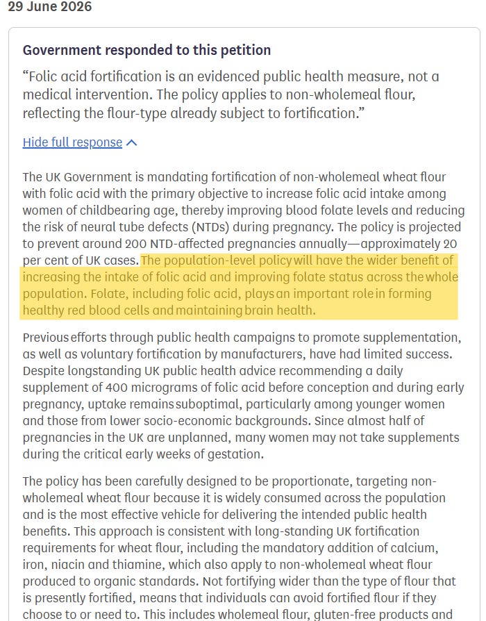
This review was prompted by a series of Substack posts by Dr Clare Craig and Hart, in response to the UK government’s planned mandatory fortification (i.e., mass medication) of non-wholemeal wheat flour from December ‘26.
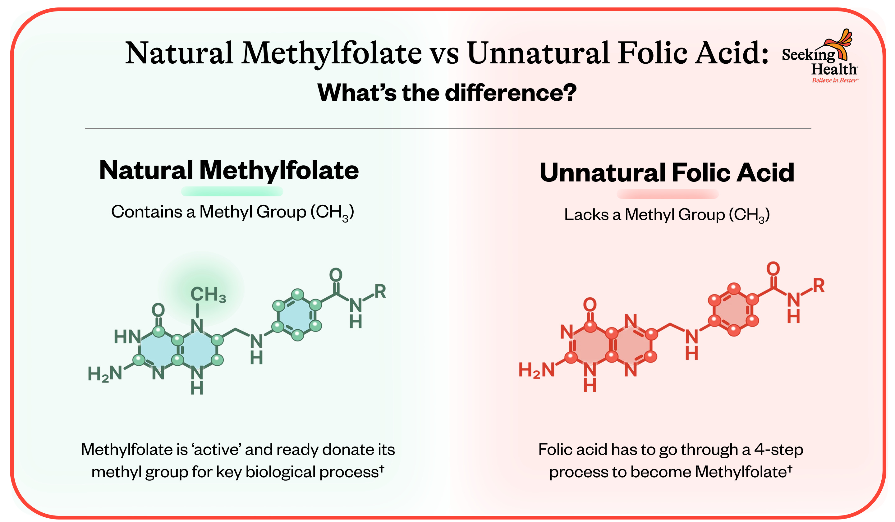
Synthetic folic acid (a synthetic form of vitamin B9) added to flour is not chemically or biologically the same as natural folate (methylfolate). The body can convert both synthetic and natural B9 into the active form, but the oxidised, synthetic form requires a slower liver-conversion process.
This is made worse for the 40-50% us which possess mutations in the MTHFR (methylenetetrahydrofolate reductase) enzyme gene—especially variants like C677T and A1298C—that reduce the enzyme’s ability to convert it into active L-methylfolate (5-MTHF) form.1
Documented risks & damage caused by excessive intake of synthetic folic acid (>1 mg/day) in individuals with MTHFR polymorphisms include:
Masking Vitamin B12 deficiency, allowing neurological damage to progress undetected.
Elevated plasma homocysteine levels, linked to cardiovascular strain, endothelial dysfunction, and thrombotic (blood-clotting) risk.
Ironically, reproductive and developmental impacts. These include recurrent miscarriages, impaired sperm/embryo viability, and increased risks of neurodevelopmental issues or autism traits in offspring.2 These can more than offset NTD cases prevented by folic acid fortification.
Rates of neural tube defects (NTDs) like spina bifida and anencephaly were already declining in several regions, including the United States and California, even before mandatory folic acid fortification was introduced in 1998.
The literature and clinical data suggest that NTDs and bottlenecks in folate availability for the developing child were, in most modern cases, not due to poor diet, but to other factors, including arsenic and mercury intake, vitamin A deficiency, and genetic factors.
Created by Dr Clare Craig Closes on 8 December 2026
We call on the Government to stop the fortification of flour with synthetic folic acid. Folic acid is a medicine not a food. We believe it is unethical to mass medicate the entire population without their knowledge or consent in the hope that a minority group of people might see a benefit.
The NHS website lists sections of the population (including people with cancer, a stent or low vitamin B12 levels) who should avoid folic acid exposure because of possible detrimental health impacts. Even if these people are aware of the issue, it is becoming harder to avoid as wholemeal and gluten free products are voluntarily adding folic acid.
What particularly piqued my interest were reports of a positive clinical signal for colorectal cancer rates, correlating with mandatory fortification and measured serum or red blood cell (RBC) folate levels.
I’m aware from my other research that a substance doesn’t need to be classed as carcinogenic (genotoxic/DNA-damaging) to promote cancer. As per the multi-hit theory of cancer, a substance that inhibits tumour suppressors can lead to similar outcomes, but not by initiating cancers, but by being conducive to the proliferation of pre-existing neoplastic lesions.
From this standpoint, I reviewed the literature to see whether synthetic folic acid affected any tumour suppressors, positively or negatively, and what mechanisms were involved. This Substack presents my findings.
Tumor suppressor genes are important genes that act within the genome to regulate several cellular functions. These genes can be broadly classified based on their role in cell growth/cell cycle progression, cell proliferation, DNA repair mechanisms, and other crucial cellular signaling functions such as the apoptosis induction.
Without functional tumor suppressor genes, there is a high risk of dysregulated cell growth that is a well-known mechanism for the development of cancers. [1]
Loss of function mutations in tumor suppressor genes has been identified in many types of cancers, including ovarian, lung, colorectal, head and neck, pancreatic, uterine, breast, and bladder cancer.[2][3][4][5][6]
… Tumor suppressor genes function to either repress or inhibit the cell cycle or promote apoptosis. The more specific functions of tumor suppressor proteins fall into several categories, including[1][21]:
Inhibition of mitogenic signaling pathways
Inhibition of cell cycle progression
Inhibition of “pro-growth” programs of metabolism and angiogenesis
My emphasis is in bold, and I lightly reformatted some passages for legibility.
2.1 Unheeded warnings
Warnings have been shouted from the rooftops since mandatory fortification began.
From “Science-based micronutrient fortification: which nutrients, how much, and how to know?“, (August, 2005), by Irwin:3
Despite a great deal of effective, although at times discordant, interaction between the 3 sections of the Department of Health and Human Services (FDA, Centers for Disease Control and Prevention, and National Institutes of Health), there was, at the time the decision was made to mandate the addition of folic acid to enriched flour, no coherent plan to monitor the national and regional effect on neural tube defect births, to document actual changes in folate status, or to assess the possible occurrence of untoward effects on the population.
… The synthesis of folic acid by Lederle Labs in 1947 was one of the milestones achieved during the era of discovery of vitamins in the first half of the 20th century. This stable and unreduced form of folate has served wonderfully in preventing and treating folate deficiency and for much of the study of folate biology.
However, folic acid is not the natural form of the vitamin as it exists in food, and evidence suggests that concentrations of unmetabolized folic acid in the blood after the ingestion of supplements or fortified foods may have different effects on folate binding proteins and transporters (9).
The tolerable upper level of folic acid intake, set forth recently by the Institute of Medicine in the Dietary Reference Intakes (10), warrants continuing consideration, especially the publication’s emphasis on the possible adverse effects of high folic acid intakes in persons with a poor vitamin B-12 status.
… Policy decision making in any country considering fortification with one or several micronutrients should not be limited to the documentation of the indications for fortification, the food or foods used to deliver the nutrients, the expected bioavailability of the nutrient, or even the dose of the fortificant.
A plan for evaluating the effectiveness and safety of the intervention is also needed. Although such an evaluation may be costly, the target population of a nationwide or global intervention deserves to be informed about its effectiveness with regard to health promotion, disease prevention, and safety.
Nutrition scientists have a major role to play in designing these evaluative protocols. The agencies responsible for national or international implementation of food fortification programs must be ready to accept the responsibility and to provide the funds for these evaluations.
The rollout in the USA, and many other countries, could only be described as reckless, if not criminally negligent.
2.2 Mechanisms
During pregnancy, folic acid levels need to be tightly regulated to prevent preeclampsia. Folic acid promotes cellular proliferation, migration and invasion of mononuclear trophoblast cells.
When you adulterate the diet of 70 million people in the UK to reduce the risk of NTDs in a couple of hundred possible cases, the risk-to-benefit ratio is hugely skewed towards unnecessary risk, as you can also promote the same proliferative pathways in pre-cancerous lesions due to off-target effects.
Methylation
Folic acid induces epigenetic changes in a cell; in other words, it can change the way genes work without changing the underlying DNA sequence.
Methylation is the key epigenetic mechanism used by folic acid:
It supplies carbon and methyl groups through one-carbon metabolism. This fuels the cellular creation of S-adenosylmethionine (SAM).
SAM is a universal methyl donor, which modifies DNA and histones.
SAM-mediated methylation inhibits the expression of some tumour suppressors.
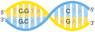
CpG methylation is an epigenetic mechanism in which a methyl group is added to the 5th carbon atom of a cytosine ring, creating 5-methylcytosine (5mC), and it occurs specifically at CpG sites (where a cytosine nucleotide is directly followed by a guanine nucleotide on the same DNA strand).
CpG methylation represses gene expression mainly by preventing transcription factors from binding to gene promoters and by recruiting proteins that condense chromatin into a closed, inactive state.
“a CpG site, i.e., the " 5'—C—phosphate—G—3' " sequence of nucleotides, is indicated on one DNA strand (in yellow). On the reverse DNA strand (in blue), the complementary 5'—CpG—3' site is shown. A C-G base-pairing between the two DNA strands is also indicated (right)” By Helixitta - Own work, CC BY-SA 4.0, https://commons.wikimedia.org/w/index.php?curid=47075331
Cases of Autism spectrum disorder (ASD) have been associated with excess or very low levels of maternal or dietary folic acid, and there is a mechanistic link between CpG methylation and neurological disorders.
The most devastating of these is the rare, progressive, and fatal neurodegenerative disorder Amyotrophic lateral sclerosis (ALS), which is also known as Lou Gehrig’s disease or motor neuron disease:
DNA methylation is a major epigenetic mark with important roles in genetic regulation. Methylated cytosines are found primarily at CpG dinucleotides, but are also found at non-CpG sites (CpA, CpT, and CpC).
The general functions of CpG and non-CpG methylation include gene silencing or activation depending on the methylated regions. CpG and non-CpG methylation are found throughout the whole genome, including repetitive sequences, enhancers, promoters, and gene bodies.
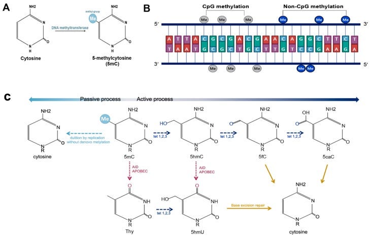
DNA methylation and demethylation. (A) DNA methylation occurs at the fifth carbon of cytosine and leads to the formation of 5-methylcytosine (5mC); (B) DNA methylation is predominantly found at CpG sites, and is much less commonly observed at non-CpG sites, such as CpA, CpT, and CpC; and (C) 5mC can be demethylated by passive or active processes. Active DNA demethylation can occur either via oxidation or deamination. The oxidation process is carried out by Ten-Eleven-Translocation (TET) proteins, including TET1, TET2, and TET3. TETs convert 5mC into 5-hydroxymethylcytosine (5hmC), which is further changed into 5-formylcytosine (5fC) and 5-carboxylcytosine (5caC). 5caC is excised and replaced via base excision repair. 5mC and 5hmC can also be demethylated via deamination by activation induced cytidine deaminase (AID)/apolipoprotein B mRNA editing enzyme, catalytic polypeptide (APOBEC).
DNA methylation plays an important role in gene regulation. It is thus not strange that some diseases are caused by abnormal DNA methylation [98]. Mutations in genes coding proteins that recognize DNA methylation or those that are required for normal neural development cause epigenetic disorders.
These disorders include RTT, fragile X syndrome (FXS), Rubinstein-Taybi syndrome, Coffin-Lowry syndrome, and alpha-thalassemia mental retardation syndrome.
In some cases, changes in histone modification or DNA methylation (hypermethylation or hypomethylation) can cause neurodegenerative disorders, such as Parkinson’s disease (PD), AD, and Huntington’s disease [99,100].
… Amyotrophic lateral sclerosis (ALS) is a neurodegenerative disease that involves motor neuron degeneration in the brain, brainstem, and spinal cord [144,145,146]. It is characterized by muscle abnormalities, including muscle spasticity, cramps, atrophy and fasciculation [147,148].
Most cases of ALS are sporadic and do not have clear causes. Aberrant DNA methylation patterns and epigenetic modifications are involved in neurodegenerative diseases [149].
Recent studies suggest that altered 5mC or 5 hmC can affect neurodegeneration, which may be caused by abnormal function of DNMTs [150,151].
Motor neurons in patients with ALS especially have changes in Dnmt1, Dmnt3a, and 5mC levels. 5mC accumulates in nuclei of motor neurons in the spinal cord and motor cortex when Dnmt1 and Dnmt3a are increased in spinal and cortical motor neurons [150].
… Although 5hmC is likely to be a stable epigenetic marker and is abundant in the adult brain, the exact mechanistic involvement of 5hmC with neurodevelopmental and neurodegenerative diseases remains unclear.
However, many researchers have suggested a potential role of 5hmC in neurological diseases, such as Rett syndrome, autism, Huntington’s disease (HD), and Alzheimer’s disease (AD) [92].
Two subsequent derivatives of 5hmC—5fC and 5caC—also transiently accumulate during neuronal and glial differentiation, but their roles in neural differentiation and neurological disease have not been studied much thus far [93].
“CpG and Non-CpG Methylation in Epigenetic Gene Regulation and Brain Function” (2017)
A paper by Rahat et al. provides some fascinating insights, with chilling implications:
“Folic Acid Levels During Pregnancy Regulate Trophoblast Invasive Behavior and the Possible Development of Preeclampsia” (2022) 4
… One of the unique features of placentation is its similarity to tumorigenesis yet being very well regulated. It allows rapid proliferation, migration, and invasion of mononuclear trophoblast cells into the maternal uterus and remodeling the maternal vasculature. This pseudomalignant nature of trophoblastic cells is strictly regulated and its importance becomes evident in abnormal pregnancies that are characterized by aberrant trophoblast proliferation/invasion like preeclampsia.
Hallmarks of high-grade neoplasms: Proliferation, invasion, metastasis, tumour cell immortality:
… Folic acid supplementation was found to increase the invasive potential of placental trophoblasts by almost two folds which were associated with the decreased expression of tumor suppressor genes and tissue inhibitors of matrix metalloproteinases; and increased expression of oncogenes, telomerase gene, and matrix metalloproteinases. These folic acid-mediated changes were observed to be regulated by CpG methylation in the case of many genes.
… Our previous study has reported the role of lower folate levels in the development of preeclampsia (22). A 2018 meta-analysis has also related folate supplementation with reduced risk of preeclampsia (23). Hence, we designed this study to analyze the effect of folic acid levels on the different epigenetic phenomena during different trimesters in normal pregnancy and their role in the development of preeclampsia.
Effect of Folic Acid Supplementation on Gene-Specific Promoter Methylation
We studied the effect of folic acid supplementation on the gene promoter region methylation in placental cell lines, viz JEG-3 and HTR-8/SVneo cells. For this purpose, we selected various categories of genes that are known to play role in regulating pseudomalignant/invasive nature of the placenta, based on the available literature and our previous studies.
The various types of the genes that we studied were tumor suppressor genes (RASSF1A, P16, RB1, PRKCDBP, APC), oncogenes (c-myc, c-jun, VEGF, EGFR), telomerase (hTERT), and matrix metalloproteinases and their inhibitors (MMP2, MMP9, TIMP1, TIMP2).
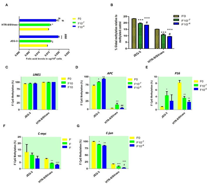
Among the tumor suppressor genes, folic acid supplementation induced a significant change in promoter methylation of APC and P16. Promoter methylation was found to be significantly increased at APC promoters with the folic acid treatment increasing in JEG-3 cells by 1.14 fold (p < 0.05) and 1.28 fold (p < 0.01) at 10−7M and 10−4M folic acid treatment, respectively, compared to untreated control cells, while in HTR-8/SVneo cell APC methylation increased significantly by 4.1-fold at 10−7M only (Figure 5D).
Inhibition of P16 expression by folic acid matters because P16 (p16INK4A) is a critical tumour suppressor gene that controls cell cycle progression, and hypermethylation effectively blocks transcription machinery from binding, downregulating and epigenetically silencing P16 expression.
The literature shows that hypermethylation of the p16 promoter drives several major epithelial cancers.
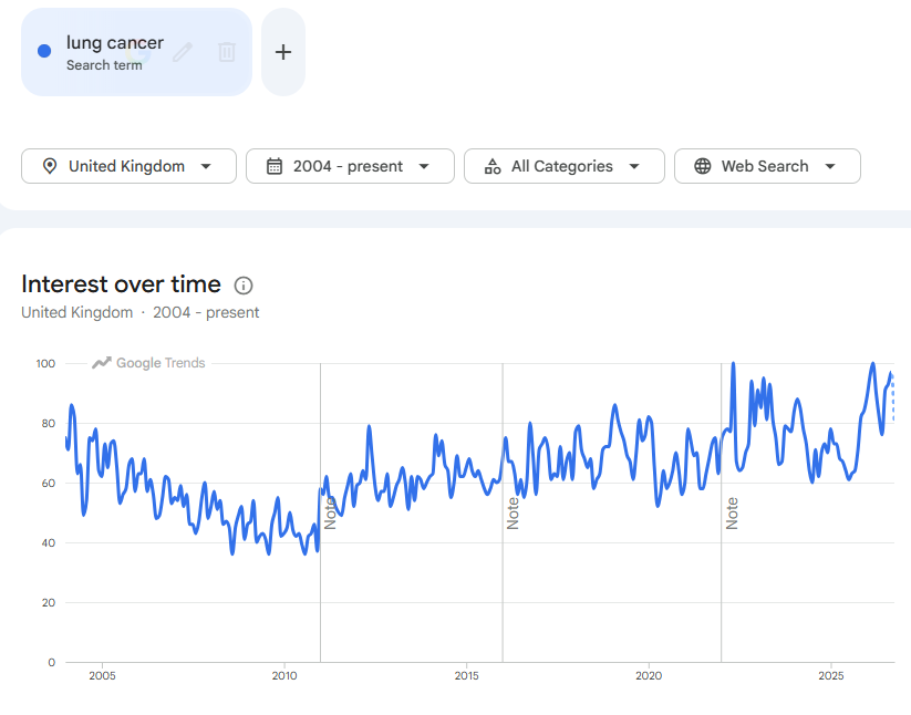
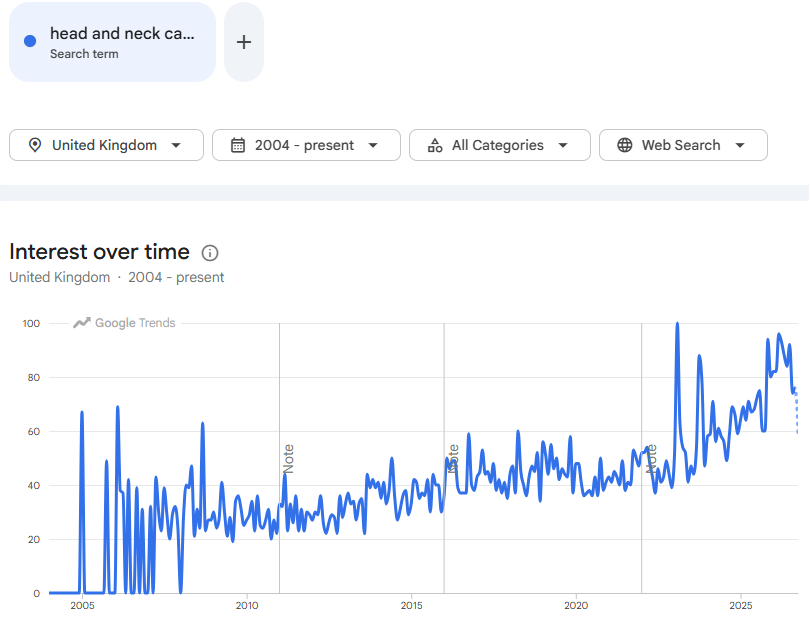
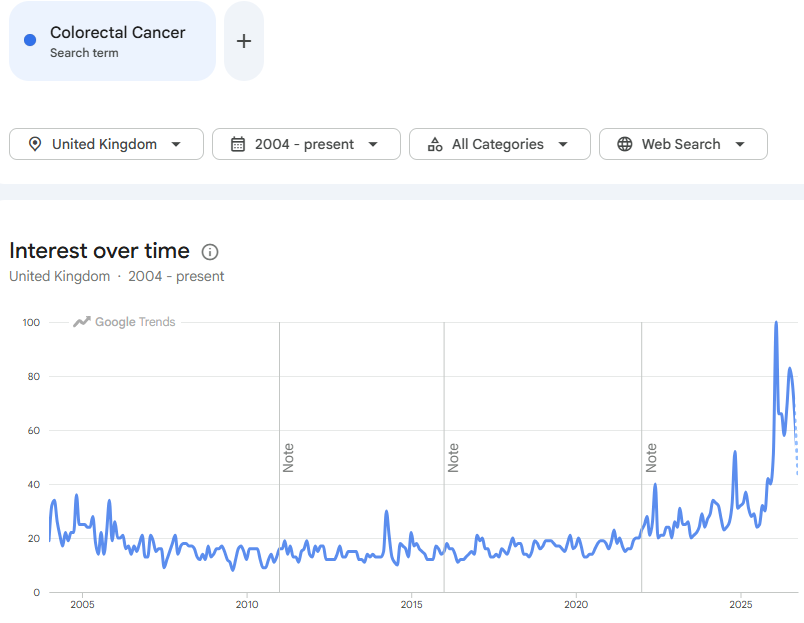
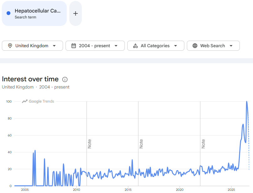
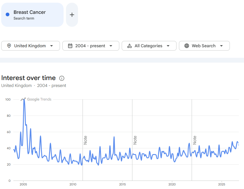
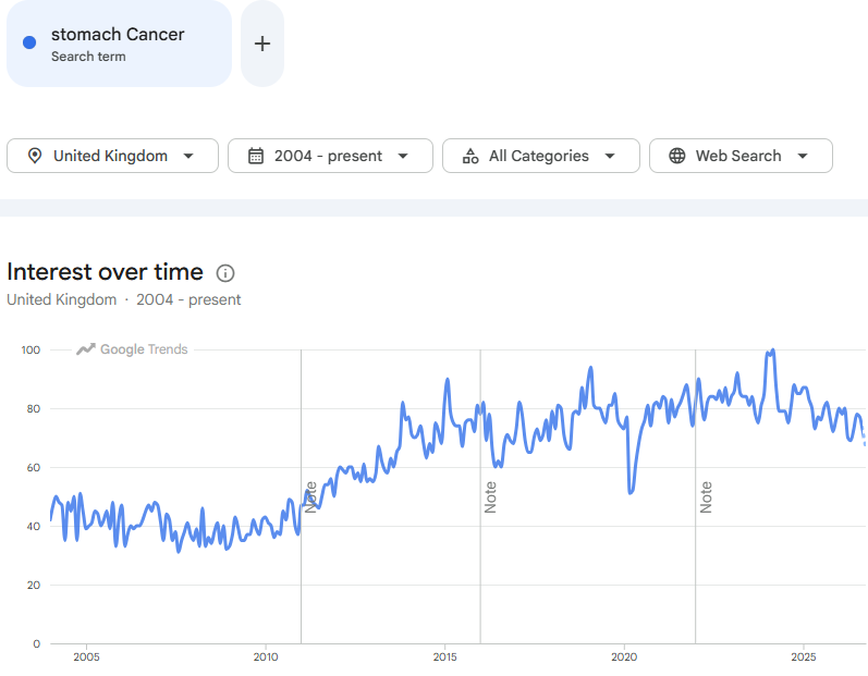
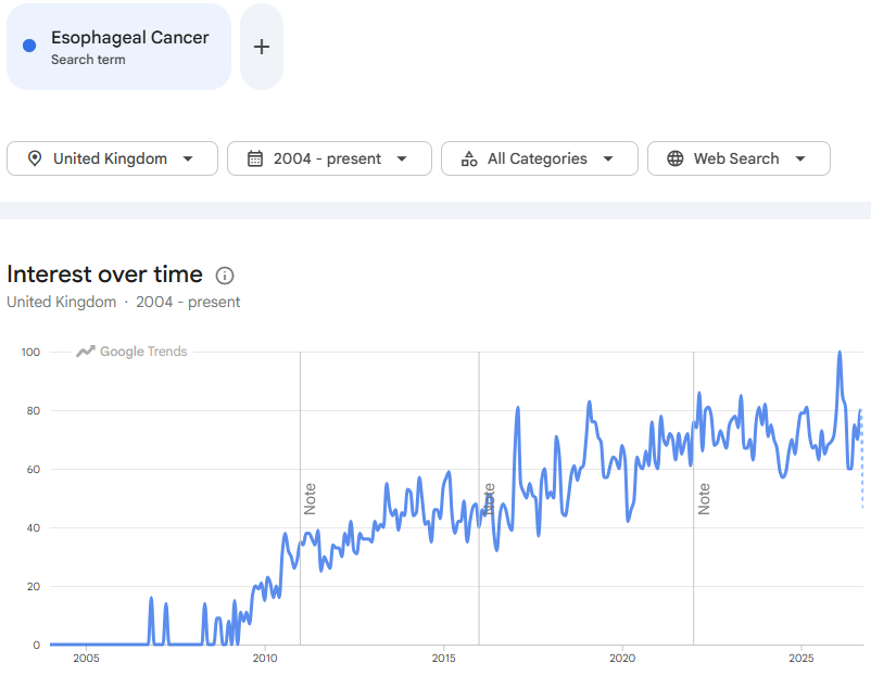
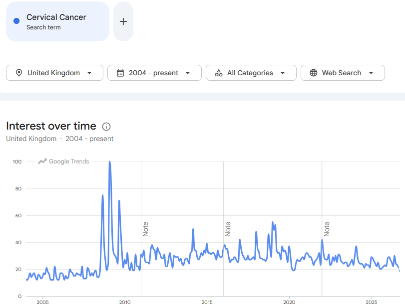
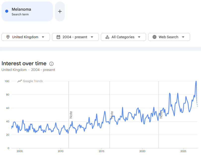
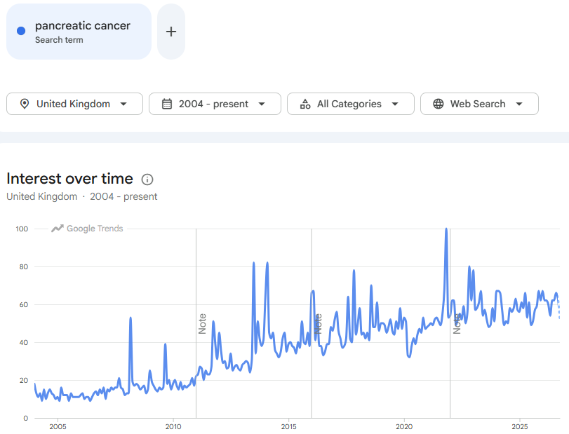
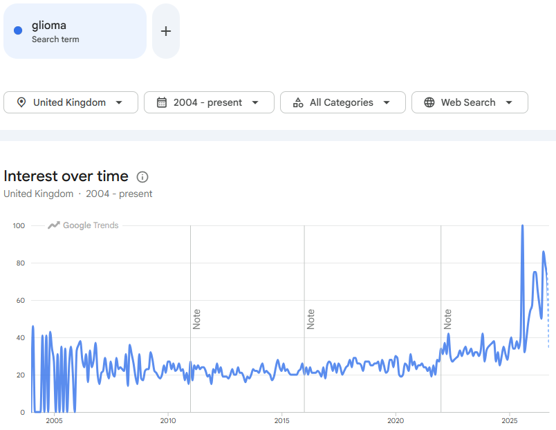
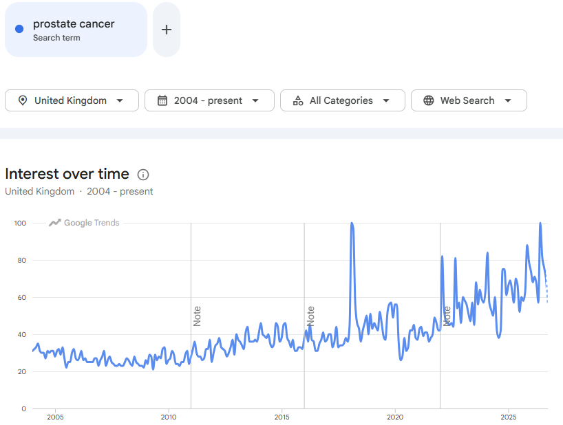
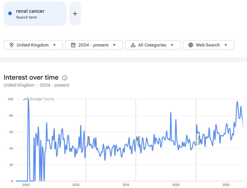
I’ve added Google Trends search interest data from 2004, where available.
These are only indicative, but some are quite concerning, and most trends are heading in the wrong direction.
Non-Small Cell Lung Cancer (NSCLC).
Head and Neck Squamous Cell Carcinomas (HNSCC).
Colorectal Cancer (CRC).
Hepatocellular Carcinoma (HCC).
Breast Cancer.
Gastric and Esophageal Cancers.
Cervical Squamous Cell Carcinoma.
And normal P16 activity suppresses many types of cancer, including:
Melanoma.
Pancreatic Adenocarcinoma.
Gliomas.
Prostate and Renal Cancers.
If you want to look further for clinical signals linked to excess dietary folic acid, I would start here. This Substack later reviews papers which indeed indicate this for CRC and prostate cancers.
“Surprisingly” - one of my favourite words to see in a research paper:
Surprisingly, folic acid-modified the P16 promoter methylation (Figure 5E) in a reverse manner between choriocarcinoma cell line, JEG-3, and normal first- trimester cell line HTR-8/SVneo. P16 promoter methylation increased in JEG-3 cells by 5 fold (p < 0.05) at 10−7M folic acid treatment compared to untreated cells, while in HTR-8/SVneo cells folic acid was found to decrease the P16 methylation by 2.4 fold (p < 0.01) at 10−7M folic acid treatment and by 3.5 fold (p < 0.01) at 10−4M folic acid treatment.
Figure 5.
Effect of folic acid supplementation on DNA methylation in JEG-3 and HTR-8/Svneo cell lines. (A) Relative folic acid levels after folic acid supplementation (B) Percentage global methylation upon folic acid supplementation (C–G) Effect of folic acid supplementation on 5'CpG Methylation (%) in (C)LINE1 promoter region, (D)APC promoter region, (E)P16 promoter region, (F)c-myc promoter region, and (G)c-jun promoter region. F0- without folic acid supplementation, F10−7and F10−4 - folic acid supplementation at the concentration of F10−7 and F10−4 M respectively. The data are expressed as mean ±SEM of three experiments. *p < 0.05, **p < 0.01, ***p < 0.001 with respect to the F0 control and #p < 0.05, ##p < 0.01, ###p < 0.001 with respect to the F10−7 treated cells.
Folic Acid Supplementation Alters mRNA Expression in Trophoblast Cells
Further to study the transcriptional profile of various genes under study, upon folic acid supplementation, we quantified the mRNA levels of various studied genes in JEG-3 and HTR-8/SVneo cell lines after giving folic acid treatment at 10−7 and 10−4 M.
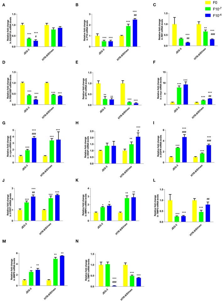
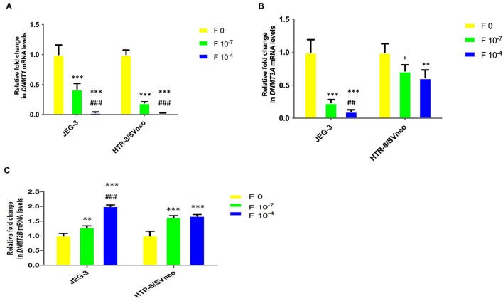
This paper found that excess folic acid can inhibit at least 4 different tumour suppressors and, as before, promotes oncogenes, MMPs (linked to tissue invasion), and decreases levels of DNA methyltransferase 1 (DNMT1).
Lower levels of DNMT are linked to neurological and neurodegenerative diseases through disruptions in gene expression, chromatin stability, and cellular homeostasis
Folic acid treatment was found to significantly decrease the mRNA levels of studied tumor suppressor genes, RASSF1A, RB1, PRKCDBP, and APC either in one or both cell lines in a dose-dependent manner. However, folic acid was found to induce a reverse trend in mRNA levels in JEG-3 and HTR-8/SVneo cells in the case of P16 as shown in Figures 6A–E.
Figure 6.
Effect of folic acid supplementation on mRNA levels of specific genes in JEG-3 and HTR-8/Svneo cell lines. Relative fold in mRNA levels of levels of (A)RASSF1A(B)P16(C)RB1(D)PRKCDBP(E)APC(F)c-myc(G)c-jun(H)VEGF(I)EGFR(J)hTERT(K)MMP2(L)TIMP2(M)MMP9(N)TIMP1 F0- without folic acid supplementation, F10−7and F10−4 - folic acid supplementation at the concentration of F10−7 and F10−4 M, respectively. The data are expressed as mean ±SEM of three experiments. *p < 0.05, **p < 0.01, ***p < 0.001 with respect to the F0 control and #p < 0.05, ##p < 0.01, ###p < 0.001 with respect to the F10−7 treated cells.
“hTERT expression” refers to the production of messenger RNA (mRNA) and protein from the human telomerase reverse transcriptase (hTERT) gene, which encodes the core catalytic subunit of the telomerase enzyme complex, which is linked to cancer cellular immortality, despite repeated cell division cycles.
Folate was found to act as an inducer of studied oncogenes and hTERT expression. The expression of c-jun, c-myc, EGFR, and hTERT increased in a dose-dependent manner in both cell lines, while only HTR-8/SVneo cells showed such dose-dependent increased expression for VEGF (Figures 6F–J).
Folic acid supplementation altered the expression levels of MMPs and their inhibitors in a reverse trend in both cell lines. The expression of MMP2 and MMP9 increased, while that of TIMP1 and TIMP2 decreased in a dose-dependent manner in JEG-3 and HTR-8/SVneo cells (Figures 6K–N).
Folic Acid Supplementation Alters mRNA Expression of DNMTs
DNMT1 mRNA level in placental cell lines showed a significant decrease upon the folic acid supplementation (Figure 7A). The decrease in DNMT1 expression at 10−7M folic acid treatment was 2.3- and 5-fold in JEG-3 and HTR-8/SVneo cells, relative to their respective control cells. It was further reduced by 11.8-and 7.8-fold at 10−4M folic acid treatment, with respect to their respective 10−7M folic acid-treated cells.
Figure 7.
Relative fold change in mRNA levels of DNMTs upon folic acidsupplementation in JEG-3 and HTR-8/SVneo cells. (A) DNMT1, (B) DNMT3A, and (C) DNMT3B. F0- without folic acid supplementation, F10−7and F10−4 - folic acid supplementation at the concentration of F10−7 and F10−4 M, respectively. The data are expressed as mean fold change ±SEM of three experiments. *p < 0.05, **p < 0.01, ***p < 0.001 with respect to the respective F0 control and ##p < 0.01 and ###p < 0.001 with respect to respective F10-7 treated cells.
A Cochrane review and the authors caution against unregulated supplementation with folic acid during pregnancy:
Folic acid was found to induce a dose-dependent effect on the invasive potential of trophoblast cells. Folic acid supplementation at 10−7 M was found to increase the invasive potential of trophoblasts by more than 2-fold, showing invasion enhancing effect of folic acid at physiological concentration.
A dose-dependent increase in invasive potential has earlier also been observed in extravillous trophoblasts from first-trimester placental explants at folic acid concentrations of 10−6 M, 10−8 M, and 10−10 M which represent physiological ranges (17).
Furthermore, treatment with the anti-folate drug methotrexate in ectopic pregnancy has been shown to reduce placental growth and trophoblast invasion due to reduced trophoblast proliferation (46).
Thus, we propose that at physiological concentrations, folic acid is able to increase trophoblast invasion, and therefore, play a key role in the regulation of trophoblast invasion, which is a crucial part of placental development, while at very high concentration, it reduces the invasion drastically.
This biphasic nature of the response to folic acid supplementation, therefore, emphasizes the importance of careful evaluation of concentration before implementation of supplementation during pregnancy.
This issue was also highlighted in a Cochrane review of micronutrient supplementation in pregnancy (47). Concentrations higher than the current daily recommendation of 400 μg may be harmful to placental development.
Specific treatment with excess folic acid also results in reduced cellular viability in the villous trophoblast (18). Although too little folic acid results in nervous tissue damage, as is accepted by the scientific community in regard to neural tube defects, a massive dose of folic acid in utero has been correlated with the development of autism in children (48).
From a 2020 study by Tong et al., “MiR-22, regulated by MeCP2, suppresses gastric cancer cell proliferation by inducing a deficiency in endogenous S-adenosylmethionine”,5 we learn that excess folic acid can, via SAM, inhibit the expression of several microRNAs, including MiR-22, which potently suppresses gastric cancer (GC) cells:
Abstract
This study investigated the effect of methyl-CpG-binding protein 2 (MeCP2) on miRNA transcription. Our results of miRNA chip assay and ChIP-seq showed that MeCP2 inhibited the expressions of numerous miRNAs by binding to their upstream elements, including not only the promoter but also the distal enhancer.
Among the affected miRNAs, miR-22 was identified to remarkably suppress gastric cancer (GC) cell proliferation, arrest G1–S cell cycle transition, and induce cell apoptosis by targeting MeCP2, MTHFD2, and MTHFR.
Understanding GC metabolism characteristics is the key to developing novel therapies that target GC metabolic pathways. Our study revealed that the metabolic profiles in GC tissues were altered.
SAM (S-adenosylmethionine), a universal methyl donor for histone and DNA methylation, which is specifically involved in the epigenetic maintenance of cancer cells, was found increased.
The production of SAM is promoted by the folate cycle. Knockdown of MTHFD2 and MTHFR, two key enzymes in folate metabolism and methyl donor SAM production, significantly suppressed GC cell proliferation.
MiR-22 overexpression reduced the level of endogenous SAM by suppressing MTHFD2 and MTHFR, inducing P16, PTEN, and RASSF1A hypomethylation.
In conclusion, our study suggests that miR-22 was inhibited by MeCP2, resulting in deficiency of endogenous SAM, and ultimately leading to tumor suppressor dysregulation.
The MTHFR gene encodes the enzyme methylenetetrahydrofolate reductase, which we discussed earlier in the context of MTHFR polymorphisms.
The MTHFR enzyme converts 5,10-methylenetetrahydrofolate into 5-methyltetrahydrofolate (active folate or L-methylfolate), and knockdown of MTHFR also suppresses gastric cancer cell growth:
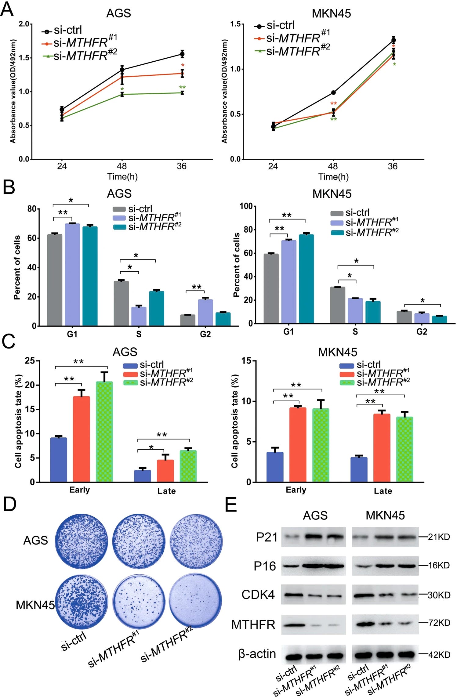
To explore whether MTHFR dysregulation affects GC cell growth, we transfected the MTHFR siRNA to AGS and MKN45 cells. Through cell viability, flow cytometry, and clone formation assays, we found that MTHFR silencing could suppress GC cell proliferation and cell cycle G1/S phase transformation and induce cell apoptosis (Fig. 4a–d and Supplementary Fig. S4).
To analyze the molecular mechanism of MTHFR, western blot analysis was used to detect the effect of MTHFR silencing on downstream proteins. The results showed that MTHFR affected GC cell growth by regulating the cell cycle-related proteins, such as P21, P16, and CDK4 (Fig. 4e).
si-MTHFR: MTHFR knockdown caused by small interfering RNA (siRNA).
Panels A & D show that si-MTHFR slows cell proliferation relative to the control (si-ctrl), and fewer colonies form.Panel
B shows that cells arrest, or become trapped, primarily in the G1 phase of the cell cycle, preventing effective replication.
C shows how apoptosis rates of i-MTHFR groups (Orange and Green bars) climb dramatically in both Early and Late stages for both cell lines (AGS and MKN45), relative to si-ctrl control (blue bars, very low levels of apoptosis in the 3-9% range):
Fig. 4: MTHFR promotes GC cell proliferation.
a MTT assay of cell proliferation at 24, 48, and 72 h after transfection with MTHFR siRNAs. b Flow cytometry of cell cycle, visualized by PI staining. c Cell apoptosis by flow cytometry, visualized using Annexin-V/PI staining. d Colony formation assay 14 days after transfection. e Effect of MTHFR knockdown on cell cycle-related proteins. All the results are shown as mean±SD. n = 3, *p < 0.05, **p < 0.01, Student’s t test.
p21 protein (p21Cip1/Waf1, encoded by the CDKN1A gene) acts as tumour suppressor:
… Studies have also shown that low doses (0.1.1 mM) of SAM promote colon cancer cell line growth and inhibits proliferation at high doses (3 mM)49. At the same time, dietary intake of high concentrations of folic acid, an important molecule in carbon metabolism, can increase levels of serum folic acid, which can lead to DNA methylation of P16, MLH1, and MGMT50.
Our study demonstrates that SAM reduces the expression of the tumor suppressor genes P16, P21, PTEN, and RASSF1A. Interestingly, P21 inhibition is mainly regulated by histone methylation51,52. However, this does not contradict our conclusion that SAM deficit induced P21 upregulation.
… In conclusion, miR-22 expression is inhibited by MeCP2. MiR-22 targets MTHFD2 and MTHFR to inhibit SAM synthesis and induces P16, P21, PTEN, and RASSF1A upregulation, which inhibits the proliferation of GC cells. This study suggests that the MeCP2 -miR-22-MTHFD2-MTHFR axis may be a therapeutic target for GC patients.
“Association of Folate and Vitamins Involved in the 1-Carbon Cycle with Polymorphisms in the Methylenetetrahydrofolate Reductase Gene (MTHFR) and Global DNA Methylation in Patients with Colorectal Cancer” (2019) by Ferrari et al.6 investigated correlations between serum folate, dietary folate, CRC and DNA methylation status, and cancer staging.
They found several statistically significant associations:
… The study included 189 patients with colorectal adenocarcinoma answering a clinical evaluation questionnaire and the Food Frequency Questionnaire (FFQ) validated for patients with colon and rectal cancer.
Blood samples were collected for evaluation of MTHFR gene polymorphisms in global DNA methylation in blood and in tumor.
The values for serum folate were positively correlated with the equivalent total dietary folate (total DFE) (rho = 0.51, p = 0.03) and global DNA methylation (rho = 0.20, p = 0.03).
Individuals aged over 61 years (p = 0.01) in clinicopathological staging III and IV (p = 0.01) and with + heterozygous mutated homozygous genotypes for the MTHFR A1298C gene had higher levels of global DNA methylation (p = 0.04).
The association between dietary intake of folate, serum folate, and tumor stage were predictive of global DNA methylation in patients’ blood. The levels of serum folate, the dietary folate and the status of DNA methylation can influence clinicopathological staging.
… during tumor development, studies point to two important epigenetic alterations. First, hypermethylation of promoter regions rich in CG dinucleotides lead to a silencing of tumor suppressor genes [43]. The silencing mechanism is possibly due to a lack of binding of transcription factors and changes in the chromatin structure, since the methylated DNA recruits complexes of proteins that promote the deacetylation of the histones, leading to the compaction of the chromatin.
Thus, DNA methylation represses transcription by inhibiting binding of transcription factors and by recruitment of proteins that bind to methylated CpG [41]. The silencing of tumor suppressor genes can promote the initiation of carcinogenesis and neoplastic progression by inhibition of apoptosis and autophagy pathways in cancer cells [44].
In addition, it is known that when the cytosine molecule is methylated and converted to 5-methylcytosine, it is unstable and may undergo spontaneous deamination, being converted into thymine. This type of point mutation (C-G for T-A) may influence the function of oncogenes or tumor suppressor genes [45].
Secondly, global hypomethylation of DNA is observed, interfering with genomic stability, reactivation of transposable elements, and loss of gene imprinting patterns, thus contributing to tumor development [41,46,47].
In addition to these mechanisms, some studies suggest that alterations in epigenetic mechanisms may influence the epithelial-to-mesenchymal transition, through the silencing of integrins, and consequently tumor progression.
It is also known that epigenetic mechanisms may interfere with miRNA, which are involved with tumor development and progression [48].
In the present study, patients with more advanced staging had higher levels of global DNA methylation when compared to patients in earlier stages. It is already well established that the global hypomethylation of DNA is a frequent and early characteristic of carcinogenesis, listed with the prognosis of several types of tumors.
… at the onset of tumorigenesis, the presence of global DNA hypomethylation that can lead to genomic instability is well established. However, as found in the present study in more advanced clinicopathological stages, one of the possible hypotheses would be the global hypermethylation of the DNA being the cause of the more advanced staging or the consequence of the same.
Dietary folate protects against some cancer types and cardiovascular disease, but it also has a dual role. If you already have pre-cancerous lesions, then excess folic acid (particularly the oxidised, synthetic form) may promote transformation, cancer growth, and disease progression:
… Clinicopathological staging and serum folate levels were predictive variables of this epigenetic event. However, no other studies were found with patients undergoing pre-treatment by colorectal cancer that evaluated these variables as being predictors of global DNA methylation.
I would like to frame this and send it to various members of parliament and public health advisory bodies:
Despite the protective action of folic acid against CCR [51], some studies call attention because of its double role, assuming that the folate intake, through an already established pre-neoplastic lesion, can accelerate the growth of tumor cells [50].
Some theories try to explain the double role of folic acid during colorectal carcinogenesis. Firstly, it is important to emphasize that adequate folate intake is associated with numerous health benefits, which encompass reduction of neural tube defects, reduced risk of cardiovascular diseases and cancer.
However, in individuals without pre- or neoplastic lesions, low folate intake and decreased serum folate levels have been associated with increased risk of colorectal tumors [51]. The mechanism by which folate deficiency can lead to colorectal cancer is related to synthesis of purines and thymidylate.
Thus, adequate levels are essential for the synthesis, stability, integrity, and adequate repair of DNA. In addition, folate deficiency is also associated with chromosomal instability, changes in transcriptional regulation, poor incorporation of the uracil into DNA, and hypomethylation of this molecule [52].
However, in cells with intense cell division, folic acid appears to act differently. First, based on the assumption that pre-neoplastic lesions or neoplastic cells have high replication and proliferation rates, high levels of folate could favor the synthesis and replication of DNA through nucleotide synthesis.
Balance and an adequate supply are key, and the optimal amount of folate you need depends on context.
A deficiency of folic acid in your diet may be beneficial if you already have cancer; therefore, a government-mandated one-size-fits-all approach to supplementation is not appropriate. Politicians aren’t physicians.
Thus, in neoplastic cells, folic acid deficiency could lead to a change in their metabolism and DNA synthesis, thus resulting in inhibition of tumor growth and even tumor regression [53].
Kim (2004), analyzing animal studies and clinical observations, focused on the double role of folate in carcinogenesis, showing that this nutrient in doses above the recommended level may increase disease progression when pre-lesion is already present.
It may even trigger recurrences:
In fact, some authors point out that folate intake above the daily nutritional recommendation in addition to not reducing the risk for colorectal cancer but also increases the risk of an individual developing this type of tumor and even the recurrence of adenomas [54].
The “money shot”:
Concern increases when reminded of the mandatory fortification of folic acid in food, which is the most important action in the field of nutrition and public health [55].
However, Wolpin et al. (2008) evaluated the serum folate of 301 patients with colorectal tumors, in order to verify if this nutrient influences the progression of these neoplasms. As in the present study, patients who consumed a greater amount of dietary folate and who drank dietary supplements regularly had higher plasma levels of folic acid [56].
The aberrant methylation of “CpG islands” can be considered an important tumor cell proliferation factor, leading to a silencing of tumor suppressor genes and molecules involved in cell differentiation.
Hypermethylation, often observed in sporadic cases of cancer, is an event which can target several genes simultaneously, some of which are responsible for tumor genesis and/or progression [57].
More than half of the colorectal cancer present hypermethylation in at least one promoter region of tumor suppressor genes (DAPK—gene associated to apoptosis linked to protein kinase; p16INK4a and p14ARF—genes involved in the cell cycle), repair genes (MGMT-O6-methylguanine-DNA methyltransferase; hMLH1-human homolog of bacterial MutL) and genes involved in progression and metastases (TIMP3, CXCL12, ID4, and IRF8) [56].
Some studies, for example, show that hypermethylation of “CpG islands” in the promoter region of the gene may lead to loss of MGMT [58]. In addition, DNA repair mechanisms can be affected according to DNA methylation status and nucleotide availability.
Thus, increased rates of folic acid may lead to hypermethylation of tumor suppressor genes and other antineoplastic genes, inactivating them, and promoting tumor progression [59].
Supplementation with high doses of folic acid is able to increase DNA methylation. A randomized study, in which 20 patients with adenomas received or not folic acid supplementation for 1 year after polypectomy found that folate supplementation increased both serum folate levels and the degree of DNA methylation, with the latter observed at 6 months for patients with supplementation and 1 year for the placebo group [60]. Other similar studies concluded that folic acid supplementation increases serum folate levels and DNA methylation [61].
Thus, epigenetic events are increasingly being studied as they do not reach the altered gene information and are therefore potentially reversible. DNA methylation is considered to be the most important epigenetic phenomenon because it is essential for normal gene expression, DNA maintenance and integrity, chromosomal modifications, as well as for the appearance of mutations. This event occurs when the DNA methyl transferase enzyme transfers a methyl grouping of the SAM molecule to cytosine residues of the “CpG islands”.
In the last decade, several animal and epidemiological studies have reported beneficial effects of folate during carcinogenesis in different organs. However, recent studies have shown concern about the double role that folic acid can play, depending on the dose and the moment that the nutrient is ingested [62].
In our study we found that folic acid intake interferes with serum folate levels. In addition, folate intake, serum folate levels, global DNA methylation and age were predictors of clinicopathological staging.
Although the present study was not designed to evaluate DNA methylation as a biomarker of metastasis, the data found here may stimulate further investigations to better understand this association and DNA methylation as a potential biomarker of tumor progression.
A high frequency of methylated genes is strongly associated with pre-cancerous polyps and colon cancer development. It’s even given its name to a new colon cancer subset: CpG island methylator phenotype (CIMP).
From “Epigenetics and Colorectal Cancer” (2011), by Lao & Grady:7
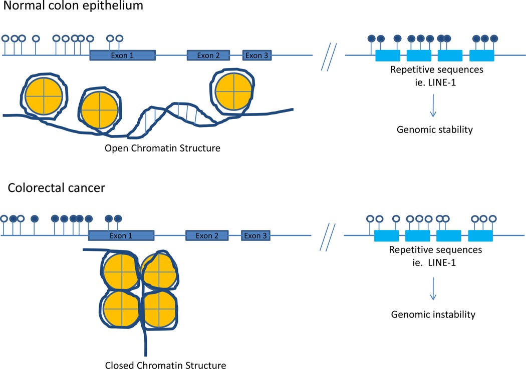
Assessment of the colon cancer epigenome has revealed that virtually all CRCs have aberrantly methylated genes and that the average CRC methylome has hundreds to thousands of abnormally methylated genes.
As with gene mutations in the cancer genome, a subset of these methylated genes, called driver genes, is presumed to have a functional role in CRC.
The assessment of methylated genes in CRCs has also revealed a unique molecular subgroup of CRCs called CpG island methylator phenotype (CIMP) cancers; these tumors have a particularly high frequency of methylated genes.
… It is now appreciated that there are multiple molecular pathways to colon cancer, and that these pathways involve both mutations and epigenetic alterations. For example, serrated polyps are associated with microsatellite instability and aberrant DNA methylation, whereas tubular adenomas more commonly arise via inactivation of the APC tumor suppressor gene and concurrent genetic alterations resulting from chromosomal instability3.
At the present time, we appreciate that there are at least three, and likely more, polyp→cancer progression sequences that arise via different molecular events.
… Epigenetic alterations, like gene mutations, contribute to the pathogenesis and molecular heterogeneity of these cancers. This is highlighted by the identification of a CpG island methylator phenotype (CIMP) subtype of colorectal cancer that has a distinct epigenome with a high frequency of methylated genes6.
Our understanding of epigenomic alterations in colorectal cancer, however, is less advanced than that of our understanding of gene mutations, but substantial progress has recently been made in this area.
Figure 1.
CpG Island DNA hypermethylation and global DNA hypomethylation in colorectal cancer as compared to normal colonic epithelium. Open lollipops represent unmethylated CpG sites in the genome. Closed lollipops represent methylated CpG sites within the genome. Unmethylated CpG islands within the promoter region of genes are correlated with an open chromatin structure (euchromatin) whereas methylated CpG islands are correlated with a condensed, closed chromatin structure (heterochromatin) and transcriptional silencing. Normal colonic epithelium generally has unmethylated CpG islands in the promoter regions of genes, whereas aberrant hypermethylation of promoter associated CpG islands is a hallmark of neoplasms. In addition to the aberrant local hypermethylation seen in colorectal cancers, global hypomethylation at LINE-1 sequences is also observed, which has been shown to associate with genomic instability. Interestingly, there is an inverse association between local CpG island hypermethylation and global LINE-1 hypomethylation as colonic neoplasms progress.
In this paper, Leubeck et al. not only modelled the effects of folic acid supplementation on rates of CRC, but they also reported a clinical signal associated with mandatory adulteration/mass medication:
“Does folic-acid supplementation prevent or promote colorectal cancer? Results from model-based predictions” (2008)8
… The bimodal role of folate in carcinogenesis is thought to be attributable to its role in nucleotide synthesis. Rapidly proliferating tissues, including tumors, have a greater requirement for folate. Cancers often overexpress folate receptors and genes in nucleotide synthesis to meet their increased need of sustained DNA synthesis and proliferation (9–11).
The irony:
Antifolate drugshave been successfully used in cancer chemotherapy and in the 1940s folate has been described as inducing an “acceleration phenomenon” if provided to children with leukemia, suggestive of increased progression (12).
The question arises whether folate intakes in the population may approach levels that could cause harm. Approximately 30–40% of adults in the United States use nutritional supplements that contain folic acid, with a standard multivitamin containing 400 μg (13).
Ironically, the group with the highest supplement use is comprised of older individuals, who are also more likely to have precancerous lesions. For example, colorectal polyps are thought to exist in about 30% of adults 60 years and older, but in many fewer younger individuals (14, 15).
In addition to supplement use, a number of functional foods are fortified with folic acid, including nutrition bars and drinks (often at 400 μg/serving), as well as fortified breakfast cereals.
Finally, the United States initiated folic acid fortification of grain products, allowable in 1996 and mandatory as of Jan 1, 1998, to reduce the incidence of neural tube defects. This public health measure has been highly effective in reducing neural-tube defects (16), yet has also increased the folic acid intake universally in the US in the population (beyond the target group of women of childbearing age) by about 100–200 μg/day (17).
Biomarkers of folate intake suggest that a significant fraction of the population has now folate levels that have been previously considered “supraphysiological”, presumably due to a combination of fortification and supplement use (17).
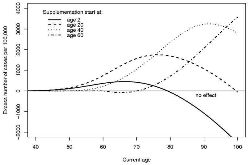
Figure 3 shows the predicted number of excess cases of colon cancer (in the absence of competing causes) as a function of current age for 4 different ages at which supplementation begins (ages 2, 20, 40 and 60) assuming a constant folate dose.
All scenarios assume that folate supplementation reduces the APC mutation rates (i.e. the rates of the first two rate-limiting events) and the malignant transformation rate by 20%, but increases the cell proliferation rate by 20% compared to (untreated) controls.
It can be seen that the cancer risk is mostly higher than the background risk (without supplementation) unless folate supplementation begins early in life.
On the other hand, it is intuitively clear that, when folate is given late in life, the effect on cumulative risk will be small as the majority of cancer cases occur before treatment begins.
Qualitatively similar curves are obtained for a 10%/10% scenario (results not shown).
The main conclusion drawn here is that the risk of colon cancer would mostly be higher than the background risk (without supplementation) unless folate supplementation is begun early in life (well before age 20).
When folate supplementation is started late in life, its effect on the cumulative cancer risk appears to be small and mostly detrimental.
Figure 3.
Predicted number of excess cases of colon cancer as a function of current age for different ages at which supplementation begins.
… Our model suggests that, in individuals who are older when supplementation is initiated, an increase in colorectal cancer rates is expected to be seen after a period of about 10 years (see Fig. 3).
These data appear consistent with recent data from the SEER cancer registries in the United States and Canada presented by Mason et al. (27). In this ecological study, shortly after the initiation of fortification, colorectal cancer rates increased in both countries resulting in an annual excess of ~4 to 6 additional CRC cases per 100,000 individuals at risk.
This abrupt increase in cancer risk may simply reflect the accelerated development and growth of a nascent malignancy into a clinically detectable or symptomatic cancer. Although our model does not explicitly describe this process (which may be considered part of the adenoma-carcinoma transition described by the last step in our model), it allows for accelerated growth of benign adenomas on the pathway to cancer (promotion).
Our model predicts an excess CRC risk that increases with time about 10 years after supplementation is initiated (Fig. 3).
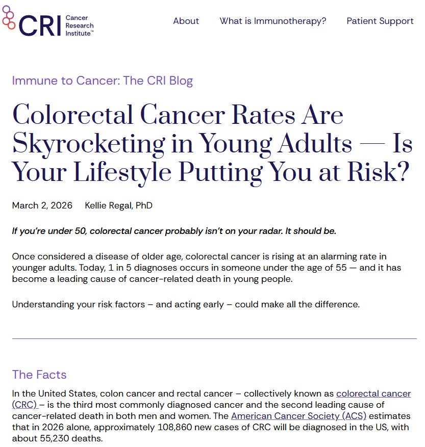
More studies are needed to see if this increase in CRC rates changes after 7 years, although headlines like this aren’t very encouraging, and the multi-hit theory will be a large part of this:
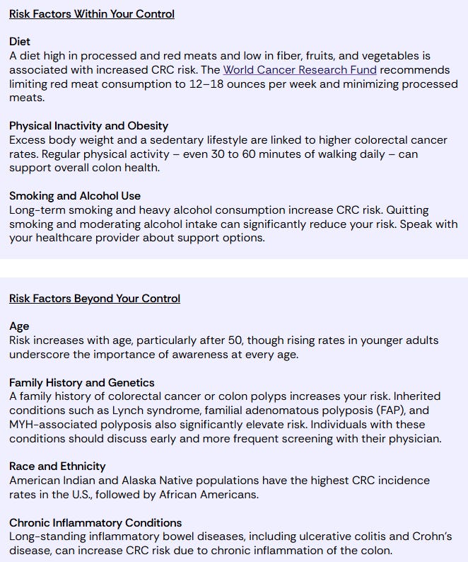
Naturally, they ignore mass administration of experimental gene therapy agents, child vaccine schedules, antibiotic misuse, the GI tract and tumour microbiome, and food adulteration with folic acid.
Note, however, Mason et al. only studied a period of 7 years (1996–2002) of folate fortification (27) and to answer the question whether or not the observed increase in CRC risk is transient, constant, or will eventually increase as predicted by our model will require more follow up data.
More warnings from the authors:
In the United States, some have suggested that a further increase in the amount of folic acid in fortified foods is warranted. Similarly, several European countries are considering the introduction of folic acid fortification for the prevention of neural tube defects.
Our results add another important piece to inform this public health policy debate. A key message is that – if excessive folate has tumor-promoting effects, then those are likely to outweigh any beneficial effects of folic acid supplementation on mutation rates with cancer rates predicted to rise in all but those treated at a very young age.
These results suggest caution when considering the implementation of fortification until we have better data on the effects of folate on cell kinetics, tumor promotion, and their quantitative effects by dose and type of folate.
I suspect the age of onset for folic acid supplementation matters less than it sounds, as hypermethylation from excess dietary folic acid is associated with inducing and promoting more cancers than it prevents.
Graphical abstract illustrating how a lifetime of excess folic acid intake balances out the negatives.
Despite possible preventative effects earlier in life, I struggle to see how a lifetime of tumour suppressor inhibition can ever be a net benefit, apart from for those in pregnancy.
Growing epidemiological and modelling evidence suggests CRC may have an extended multi-decade latency period, up to 30 years, from initiation to clinical diagnosis.9
It’s too early to give excess dietary folic acid the all-clear, and from middle age we have decades of lifespan left for potential cancers to develop.
Other mechanisms:
Excess unmetabolized folic acid (UMFA) circulating continuously through young people's peripheral blood and tissues reduces the cytotoxicity of anti-tumour Natural Killer (NK) cells.
Folic acid (FA) has different epigenetic effects depending on age. DNA repair may benefit cancer cell survival, whereas Type 3 EMT (epithelial-to-mesenchymal transition) is associated with cancer progression and metastasis. From a breast cancer study:
Comparison of the mammary transcriptome after FA supplementation in the juvenile period or adulthood revealed only nine transcripts commonly affect by FA supplementation during the two time periods.
The top pathway enriched by FA supplementation in juvenile period was DNA repair, while in adulthood it was epithelial to mesenchymal transition. FA supplementation during adulthood also led to an increase in the number of proliferating cells, increased expression of GATA3, a luminal cell marker and ductal hyperplasia.
Similar changes in OCT-4, BRCA1 and EZH2 expression were also observed in the mammary gland after feeding a FA deficient diet, with the direction of change being dependent again upon the time of exposure.
These studies show that FA intake can induce significant changes in gene expression and cell phenotype within the mammary gland, dependent upon the timing and intake of FA, with both FA deficiency and supplementation increasing the expression of genes that play a key role in mammary cell proliferation, differentiation and ductal growth, key factors implicated in breast cancer risk.
Excessive juvenile FA supplementation persistently decreases the expression of a critical pluripotency transcription factor involved in maintaining stem cell niches, OCT-4. Epigenetic or metabolic suppression of OCT-4 forces cells out of their undifferentiated stem cell state and into premature differentiation along the trophectoderm/trophoblast lineage.
It can also prematurely differentiate uncommitted stem cells needed for ongoing tissue maintenance and regeneration, leading to depletion later in life (part of premature ageing).1011121314151617
Neural Progenitor and Neural Crest Stem Cells (NCSCs / Brain: hippocampus and cerebral cortex).
Mesenchymal Stem Cells (MSCs / Bone, cartilage and fat)
Hair follicles.
Excess cellular nutrition with FA may allow damaged or mutated cells with DNA damage, or some cancer cell lines, to survive and proliferate instead of starving and dying by apoptosis.181920
One-carbon (1C) metabolism is central to the supply of nucleotides for DNA synthesis and repair. Without sufficient nucleotide supply to meet the proliferative demands, cells undergo cell cycle arrest or cell death due to replication stress and genomic instability1,2,3.
Enzymes involved in the 1C metabolism pathway are commonly upregulated in cancer to produce nucleotides and amino acids required for rapid proliferation4,5,6.
Several clinical studies have demonstrated a positive correlation between folic acid intake and prostate cancer risk,
Key takeaways from a 2018 review, “Folate and Its Impact on Cancer Risk”, by Pieroth et al.:21
Purpose of Review
Research has evaluated the potential impact of folate on cancer risk with conflicting findings. Studies have demonstrated increased risk, no effect, and decreased risk. This review summarizes findings of mixed results between folate intake, serum levels, gene polymorphisms, and cancer risk based on meta-analyses from the past five years.
Recent Finding
Low or deficient folate status is associated with increased risk of many cancers. Folic acid supplementation and higher serum levels are associated with increased risk of prostate cancer. Gene polymorphisms may impact risk in certain ethnic groups.
Two analyses also found no significant association between FA supplementation and risk of colorectal, prostate, lung, breast, or hematological cancers [32•, 33•]; however, one found a 24% increased risk of prostate cancer [34•]. One found no effect with less common types of cancer and cancers of unknown origin [33•]; and another found a 53% lower risk of melanoma [32•].
Zhang and colleagues found elevated homocysteine levels and folate deficiency, as determined by serum folate level, to be associated with increased overall risk of cancer in a meta-analysis of 83 case-control studies [11].
Folate level was inversely associated with most cancer types except prostate, bladder, pancreatic, and breast. Protection was seen in studies from Europe, Asia, the Middle East, and Latin America, but not the USA or Australia.
Two analyses found increased risk of prostate cancer when comparing highest with lowest serum folate levels. Both included five of the same nested case-control studies [17, 24].
One group included one additional case-control study and one retrospective case-control study [17]. Wang’s dose-response analysis reported a 4% increase in risk with each 5 nmol/L increase in serum folate [24].
Genotypic variation is a significant factor in your personal risk profile, and, again, the authors urge caution:
After removing a retrospective study that was contributing to significant heterogeneity, Tio’s group found the association was still significant. The study removed was conducted in Jamaica, where risk was much higher with high levels, and the authors posed the question of whether or not folate may play a role in racial differences seen in prostate cancer risk [17, 24].
In addition, more research is needed to help understand if the protective effects of folate are weaker in populations with a relatively higher intake level from mandatory fortification and/or supplementation.
Population concerns regarding fortification and increased cancer risk are challenged in the current research. Fortification status in studies did not have any impact on risk with supplementation [32•, 33•].
Fortification practices vary and increasing use of organic and gluten-free flours, which may not be fortified, may change population intake over time.
More prospective, post-fortification work needs to be done in some populations. For example, the association between supplemental intake, elevated serum levels, and increased risk of prostate cancer should be explored in the context of gene variants.
Our governments have appointed themselves as clinicians, and they certainly know better than you.
Clinicians should weigh the pros and cons of supplementation in the presence of adequate intake with individuals based on all risk factors for disease.
Finally, it is important to note that dozens of enzymes affect the efficiency of folate-related metabolic pathways, and polymorphisms in these enzymes may alter substrate availability of folate and/or other nutrients as well as gene expression.
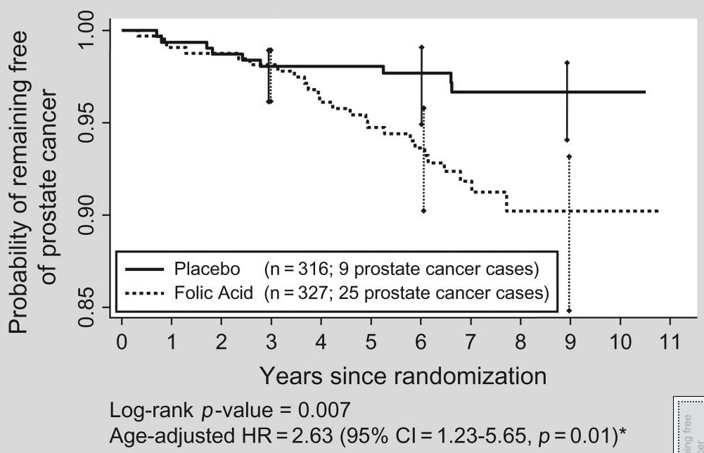
In 2009, a landmark randomised clinical trial found a statistically significant 3-fold increase in the rates of prostate cancer in men taking folic acid supplements vs aspirin or control.
From “Folic acid and risk of prostate cancer: results from a randomized clinical trial” by Figueiredo et al.:22
Abstract
Data regarding the association between folate status and risk of prostate cancer are sparse and conflicting. We studied prostate cancer occurrence in the Aspirin/Folate Polyp Prevention Study, a placebo-controlled randomized trial of aspirin and folic acid supplementation for the chemoprevention of colorectal adenomas conducted between July 6, 1994, and December 31, 2006.
Participants were followed for up to 10.8 (median = 7.0, interquartile range = 6.0-7.8) years and asked periodically to report all illnesses and hospitalizations. Aspirin alone had no statistically significant effect on prostate cancer incidence, but there were marked differences according to folic acid treatment.
Among the 643 men who were randomly assigned to placebo or supplementation with folic acid, the estimated probability of being diagnosed with prostate cancer over a 10-year period was 9.7% (95% confidence interval [CI] = 6.5% to 14.5%) in the folic acid group and 3.3% (95% CI = 1.7% to 6.4%) in the placebo group (age-adjusted hazard ratio = 2.63, 95% CI = 1.23 to 5.65, Wald test P = .01).
In contrast, baseline dietary folate intake and plasma folate in nonmultivitamin users were inversely associated with risk of prostate cancer, although these associations did not attain statistical significance in adjusted analyses.
These findings highlight the potential complex role of folate in prostate cancer and the possibly different effects of folic acid-containing supplements vs natural sources of folate.
Figure 2.
Kaplan–Meier plot of the prostate cancer–free status over time among the 643 men randomized to placebo and folic acid treatment in this study. The 95% confidence intervals (CIs) for probability of remaining free of cancer are shown at 3, 6, and 9 years for both treatment groups. The numbers of patients at risk for prostate cancer at 3, 6, and 9 years were 284, 274, and 10, respectively, in the placebo group and 300, 233, and 10 in the folic acid group. *Cox proportional hazard models were used to obtain hazard ratios (HRs) and 95% confidence intervals and Wald tests to obtain P values. In the figure, hazard ratio is age adjusted. The multiadjusted hazard ratio (with adjustment for aspirin treatment group, alcohol use, baseline multivitamin use and plasma levels of vitamins B2, B6, and B12) was 2.58 (95% CI = 1.14–5.86, P = 0.02).
According to their findings, natural, dietary folate doesn’t appear to carry the same cancer risk, for various reasons.
In fact, it appeared to reduce the risk (HR, hazard ratio) of prostate cancer by about 35% to 58%:
In contrast to the direct association of folic acid supplementation with risk of prostate cancer in the randomized analysis, there were possible indications of inverse associations of prostate cancer risk with dietary folate intake (multiadjusted HR = 0.65, 95% CI = 0.35 to 1.20), and with baseline plasma folate among subjects who did not use multivitamins (multiadjusted HR = 0.42, 95% CI = 0.17 to 1.04, Table 1). Dietary intake of B2 and B6 and plasma B2, B6, B12 was not associated with risk (data not shown).
The reason for the contrasting associations of folate supplementation and baseline intake or circulating levels of folate with the risk of prostate cancer is unknown.
Folate supplementation in the trial was in the form of folic acid, a fully oxidized, monoglutamyl form of folate that may differ in its effects from the natural reduced and methylated forms (mostly 5-methyl tetrahydrofolate).
Folic acid is more bioavailable than natural sources (20); natural folates exist as polyglutamates that are hydrolyzed to monoglutamates in the small intestine and then polyglutamated in peripheral cells (21).
The borderline inverse association of dietary intake and plasma levels (among nonmultivitamin users) is consistent with some observational data that natural folates could be protective against prostate cancer (7). However, protective dietary associations could be confounded by other factors if foods rich in folate also contain protective nutrients or are associated with protective lifestyle habits.
There are plausible biological reasons why high levels of folate may promote carcinogenesis.
Folates have a key role in the synthesis of nucleotides (1,2) that are needed for proliferating neoplastic cells, and there is an increase in the expression of folate receptors in several cancer types (22).
Prostate-specific membrane antigen, a transmembrane-carboxypeptidase with folate hydrolase activity (23,24), is overexpressed in nearly all prostate cancers, and higher tissue levels of this protein are associated with higher grade, higher Gleason score (25), and disease recurrence (26).
Folate also has a key role in DNA methylation (1,2), and alterations in methylation patterns including global DNA hypomethylation (27) and gene-specific hypomethylation (28) appear to be important events in prostate cancer.
Unmetabolized folic acid has recently been associated with reduced natural killer cell cytotoxicity (29), which could impair first-line defense against malignant cells (30).
As the policy of many governments is to deliberately make sure that there is excess folic acid in our diets, then we are left with two options:
Follow an elimination diet and eat only wheat products made with non-adulterated wholemeal flour.
Adjust your diet and review your daily stack to mitigate the effects.
You could also emigrate, but then you would need to know the history of imported food and flour, too.
The first option would work, but it would be difficult to follow, potentially expensive, and could lead to a very restricted menu, especially if you need to do all your own cooking.
For example, from this December, the following foods must all be fortified with FA:
Standard breads and baked goods: White and brown sliced loaves, rolls, baguettes, pittas, and flatbreads
Cakes, biscuits, and pastries: Commercially baked or packaged cakes, cookies, shortbread, croissants, and pastries
Pies, pasties, and sausage rolls: Products with pastry or flour-thickened components
Batter and coated products: Battered fish, chicken nuggets, and crumbed convenience foods
Puddings and sweet treats: Puddings, sweet pastries, and flour-based desserts
Certain sauces, gravies, and soups: Products that use non-wholemeal wheat flour as a thickener
Plain and self-raising white/brown wheat flour: Bags of standard non-wholemeal flour sold directly to consumers
Now, maybe we should be cutting down on many of these anyway for health reasons, but that’s a story for another day.
For practical reasons, I’ve done a literature review linked to the second option.
Repurposed & Standard Pharmaceuticals
I cannot ever advocate taking any of these just for their effects on systemic methylation, so consider this for information only:
Valproic acid, which interferes with methylation-dependent pathways and can reduce excessive methylation capacity.23 However, valproate is linked to a long list of systemic pathologies, birth defects, and a prescribing scandal. See here:
Cholesterol-lowering Bile Acid Sequestrants(Cholestyramine).24 These bind to folic acid in the GI tract, reducing bioavailability.
Anti-Folate/Folate Antagonists, such as Methotrexate or certain antimicrobials like Trimethoprim.
Oral Contraceptives.25 No single mechanism was identified in the footnoted paper, but OCs may reduce folic acid absorption; increased oestrogen levels may affect purine metabolism and thus increase folate requirements; increase urinary clearance; and increase serum folic acid-binding protein levels among OC users.
Endogenous Buffers
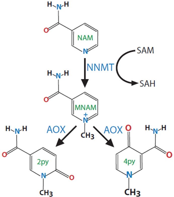
Nicotinamide(Vitamin B3) acts as a “methyl sink”, absorbing and expelling excess methyl groups to help manage elevated SAM pools.26
“Figure 1. Enzymatic pathway for NAM methylation. Nicotinamide (NAM) is methylated by nicotinamide N-methyltransferase (NNMT) using the universal methyl donor S-Adenosylmethionine (SAM). The methylated product N1-methylnicotinamide (MNAM) is further oxidized by aldehyde oxidase (Aox) into two related compounds 2py and 4py. All three metabolites are eventually excreted in the urine.” https://pmc.ncbi.nlm.nih.gov/articles/PMC5446048/
“Methylation adaptogens” are natural, plant-based compounds that inhibit DNA methyltransferases (DNMTs). SAM uses DNMTs to methylate DNA; therefore, inhibitors of these enzymes help to neutralise hypermethylation.
Examples include:
Green Tea Extract, Epigallocatechin gallate (EGCG). This well-studied natural DNMT inhibitor directly decreases global DNA methylation levels and inhibits SAM.
Curcumin has been seen to inhibit DNMTs and induce hypomethylation.
Genistein, a phytoestrogen found in soybeans, chickpeas, and other plant foods, inhibits DNMTs in a dose-dependent manner.
Resveratrol, from grape skins and red wine, has a similar effect on DNMTs as genistein.
Lycopene, found in fruits and vegetables such as tomatoes, carrots, watermelons, and papaya, has been linked to hypomethylation.
Polyphenols, including quercetin, fisetin, and myricetin. These may inhibit DNMT activity by transferring SAM to SAH.
Coffee polyphenols, including caffeic acid and chlorogenic acid, inhibit DNA methylation in a concentration-dependent manner, likely by increasing SAH formation.27
Sulforaphane (SFN), found in cruciferous vegetables, alters gene expression in the folate cycle, shifts one-carbon (1C) metabolism, and reduces DNA methyltransferase (DNMT) expression.28 The footnoted study found that SFN helps to inhibit multiple breast cancer pathways.
3.0 Conclusion
This area of folic acid fortification-associated pathologies deserves more attention than it currently receives, and I hope this review broadens the debate, so please share widely if you can.
Thanks for reading DoorlessCarp’s Scientific Literature Reviews! This post is public so feel free to share it.
I would normally advocate further research before drawing firm conclusions, but the extensive research already conducted is more than sufficient to show that the optimum amount of folic acid (preferably as folate or folinic acid) varies by genotype and medical history.
For many of us, an informed physician would not recommend supplementation, especially for those with coronary stents, due to the elevated risk of restenosis (the re-narrowing of an artery or blood vessel after a medical procedure).29
Other pathologies linked to high or excess synthetic folic acid intake discussed in the literature include:
Cancers: Colorectal, prostate and gastric cancer. There are also possible mechanisms linking excess folic acid to breast, lung, liver, cervical, pancreatic, kidney and other cancers.
Proposed mechanisms include altered DNA methylation, silencing of tumour-suppressor genes, suppression of natural killer cells, decreased expression of tissue inhibitors of matrix metalloproteinases (MMPs), increased expression of oncogenes, MMPs and telomerase, and accelerated growth of existing precancerous lesions.
Premature stem cell differentiation and depletion: A hallmark of accelerated ageing, affecting many vital organs, the brain, bones, cartilage, hair follicles, and haematology.
Vitamin B12 deficiency symptoms may be masked, potentially allowing associated neurological damage to progress undetected.
Cardiovascular and clotting problems: Elevated homocysteine levels are linked to endothelial dysfunction and increased thrombotic risk.
Pregnancy and reproductive concerns: Possible effects on placental development and trophoblast invasion, pre-eclampsia, recurrent miscarriage, and sperm or embryo viability.
Neurodevelopmental conditions: Although not shown to be caused by excess folic acid, research shows possible associations with autism-related traits, altered neurological development, and epigenetic changes in relation to conditions such as Rett syndrome, Alzheimer’s disease, Huntington’s disease and amyotrophic lateral sclerosis (ALS).
In summary, the literature shows beyond reasonable doubt that mandatory food fortification should not be implemented, and that the previously used, informed, targeted approaches to supplementation should be retained as best practice.
Via : https://www.profession-gendarme.com/. https://www.youtube.com/watch?v=_1AU30Tnojo 28 juil. 2026 VIDÉO RE-POSTÉE POUR CAUSE DE CONTENU PROTÉGÉ PAR DES DROITS D'AUTEUR Eric Mercier : infirmier D.E. dans le soin depuis 1991 (BSPP / Mitha / APHP / Libéral rural / Hémodialyse / Formateur en EHPAD). Voici son adresse de courriel : suppressionprojetloieuthanasie@gmail.com ⮕ Pour consulter le compte X d’Eric Mercier : https://x.com/Art50Mercier ⮕ La ROSP, ou Rémunération sur Objectifs de Santé Publique : https://www.ameli.fr/medecin/exercice... ⮕ "Gasping for Air", la grande étude du Dr Joël Zivot : https://www.npr.org/2020/09/21/793177... ⮕ "Gasping for Air", article sur la grande étude du Dr Joël Zivot : https://news.law.fordham.edu/blog/202... ⮕ Alicia Duncan explique le Midazolam : • ALICIA DUNCAN EXPLIQUE LE MIDA...
https://markcrispinmiller.substack.com/p/for-banning-this-essential-video? Mark Crispin Miller 31 juillet 2026 Pour avoir censuré cette vidéo essentielle en 2021, YouTube (Google) devrait être accusé de démocide, et tous ses responsables condamnés à la prison à vie sans possibilité de libération conditionnelle. Ce géant industriel, avec son credo initial « Ne soyez pas malveillants », devrait être classé au même titre qu'IG Farben, Bayer, Dow et IBM. John Leake s'exprime sur les propos psychopathes de Susan Wojcicki, PDG de YouTube, qui se vante d'avoir banni un million de vidéos qualifiées de « désinformation dangereuse » — une projection malsaine, car ces vidéos ont été bannies pour avoir révélé des vérités qui auraient sauvé d'innombrables vies. Lisez l'article inspirant de Leake, puis regardez son interview interdite avec le Dr Peter McCullough. Comme toujours, ceux — ou nous — qui avons dit haut et fort (ou essayé de dire) la terrible vérité se so...










Commentaires
Enregistrer un commentaire